Πέμπτη 27 Φεβρουαρίου 2020

Η απώλεια βάρους μειώνει τον κίνδυνο διαβήτη κατά 37%

  • Iatronet
  • 94 προβολές
    Χωρίς αξιολόγηση
Σε έρευνα, οι παχύσαρκοι με οικογενειακό ιστορικό της νόσου μπόρεσαν επίσης να περιορίσουν τον κίνδυνο διαβήτη κατά 21 % μειώνοντας τον ΔΜΣ.
Όσοι έχουν ιστορικό διαβήτη τύπου 2 μπορούν να μειώσουν τον κίνδυνο εμφάνισης της νόσου διατηρώντας υγιές βάρος, αναφέρει νέα έρευνα που δημοσιεύτηκε στο περιοδικό PLOS Medicine.
Ερευνητές ανακάλυψαν ότι μείωση κατά 1 kg/ m2 στον ΔΜΣ σε μη υπέρβαρους περιόριζε  τον κίνδυνο εμφάνισης διαβήτη κατά 37%.
Ο Manuel Rivas, δήλωσε ότι τα ευρήματα υποδεικνύουν ότι όλοι μπορούν να μειώσουν ουσιαστικά τον κίνδυνο με απώλεια βάρους.
Οι ερευνητές ενέγραψαν 300.000 Βρετανούς 40 έως 69 ετών. Σχεδόν 5% αυτών είχε διάγνωση διαβήτη τύπου 2.
Παρατήρησαν ότι σε ανθρώπους με ΔΜΣ μικρότερο από 25, που δεν ήταν υπέρβαροι και δεν είχαν οικογενειακό ιστορικό διαβήτη, μείωση κατά 1 kg/m2 οδήγησε σε 37% χαμηλότερο κίνδυνο εμφάνισης της νόσου.
Οι παχύσαρκοι με οικογενειακό ιστορικό της νόσου μπορούσαν επίσης να περιορίσουν τον κίνδυνο διαβήτη κατά 21 % μειώνοντας τον ΔΜΣ.
Αν και τα ευρήματα θεωρούνται δυνητικά σημαντικά, οι ερευνητές τονίζουν ότι χρειάζεται περισσότερη έρευνα.

Risk factors for aortic stenosis

Risk factors for aortic stenosis

Although aortic valve stenosis (AVS) shares several risk factors with atherosclerosis, lipid-lowering therapy does not reduce AVS progression. Elevated lipoprotein(a), hypertension, chronic kidney disease, and diabetes all increase the risk of AVS. In addition, recent studies have emphasised increased body mass index (BMI) as a particularly strong AVS risk factor. Modifying risk factors for AVS may reduce morbidity and potentially avoid valve interventions with health economic benefits.
Valvular Heart Disease

Introduction

Aortic valve disease includes aortic valve stenosis (AVS), aortic regurgitation (AR) and a combination of the two. The increased prevalence of non-rheumatic aortic valve disease parallels an increasingly ageing population. Degenerative AVS is the most common valvular heart disease and develops from fibrocalcific changes of the aortic valve cusps, resulting in reduced valve opening and eventually haemodynamic obstruction of the left ventricular outflow. Although a reported slight decline in AVS incidence suggested that improved cardiovascular risk factor control may limit the development of AVS in the Western world, cardiovascular prevention by means of lipid-lowering therapy has been shown to be inefficacious in reducing AVS progression. In addition to dyslipidaemia, other traditional cardiometabolic risk factors such as obesity [1-4], hypertension [3-6], and diabetes [3, 4, 6, 7], have also been shown to increase the risk of AVS in retrospective studies. Despite this shared risk factor profile and the common co-existence of atherosclerosis and valvular calcification, a substantial proportion of patients with AVS do not have concomitant coronary artery disease. Assessing the association of traditional cardiovascular risk factors with incident aortic valve disease is therefore important in order to identify potential preventive strategies in valvular heart disease. Identifying key risk factors for AVS may, in addition, provide clues for risk stratification and future interventional trials to slow down AVS progression and to avoid, or at least postpone, aortic valve interventions.

Aortic Stenosis Interventions

Over time, the number of aortic valve interventions has increased both in Europe (Figure 1A) and the USA (Figure 1B), with a decreasing proportion of surgical aortic valve replacement (SAVR) in favour of transcatheter aortic valve implantation/replacement (TAVI/TAVR) (Figure 1C, Figure 1D). Although TAVI increased earlier in European than in American populations, the proportion of surgical to transcatheter aortic valve interventions now approaches 60:40 on both sides of the Atlantic (Figure 1C, Figure 1D) [8]. The striking sex differences in Figure 1A and Figure 1B in terms of the interventional management of AVS (at least in part) also illustrates the higher AVS incidence in males compared with females [1-4]. The inflation-adjusted annual expenditure of AVS interventions in the USA doubled between 2003 and 2016 [8]. In addition to avoiding periprocedural and postprocedural risks for AVS patients, the prevention of AVS incidence would hence also be anticipated to have substantial health economic benefits.

Figure 1.  Aortic Valve Interventions Over Time in Sweden (left panels) and the USA (right panels). The upper panels show the number of aortic valve interventions in males (blue) and females (orange) per 100,000 individuals between 2003 and 2016/18. The lower panel show the proportion of surgical aortic valve replacement (SAVR; orange) and transcatheter aortic valve implantation/replacement (TAVI/TAVR; blue), respectively.
Panels A and B represent data from the Swedish National Board of Health and Welfare (Socialstyrelsen), accessed on 23/11/2019 at http://www.socialstyrelsen.se. Panels B and D from Alkhouli M, et al. Eur Heart J. 2019 [8] are reprinted by permission of Oxford University Press on behalf of the European Society of Cardiology.

208_Back_Figure 1.png


Lipids and Lipoproteins in Relation to AVS Risk

Observational studies have established an association of increased levels of low-density lipoprotein (LDL) cholesterol with AVS. Likewise, a recent Mendelian randomisation study showed a positive association between genetically predicted LDL cholesterol levels and AVS [9]. However, large clinical trials revealed a lack of effect of statin treatment to decrease the haemodynamic progression of AVS. Whether these conflicting results represent pleiotropic effects of statins of which some may be detrimental for AVS or the lack of a causal effect of LDL cholesterol on AVS remains unclear. Other lipoproteins have attracted an increasing interest for their possible causal involvement in AVS. Lp(a) is an atherogenic lipoprotein that was identified as being genome-wide significantly associated with aortic valve calcification and AVS [10]. Mechanistic studies have also generated support to the importance of Lp(a) and Lp(a)-associated oxidised phospholipids in AVS pathophysiology [11]. Furthermore, increased Lp(a) levels in patients with AVS is an indicator of faster haemodynamic progression [12] as well as increased valve calcification activity as determined using the radiotracer [13] F-NaF for PET imaging [14]. Mendelian randomisation studies have associated elevated Lp(a) levels and corresponding genotypes with increased risk of AVS in the general population, with a 10% to 30% increase in the risk of AVS per 10 mg/dL increment of genetically predicted Lp(a) levels [15]. Compared with other cardiovascular outcomes, Lp(a) lowering could potentially prevent 1 in 7 cases of AVS compared with 1 in 14 cases of myocardial infarction [16]. Importantly, statins may not alter Lp(a) levels, which should be taken into account when considering the lack of beneficial effects obtained by LDL-lowering in AVS. Although it remains to be established whether these observations translate into a therapeutic value of Lp(a) lowering for slowing down AVS progression, Lp(a) could potentially also be considered as a biomarker to guide clinical follow-up, timing of intervention, and risk stratification in AVS patients.

Obesity

We recently reported that that body mass index (BMI) is associated with the risk of developing AVS (Figure 2). In brief, our analysis involved 71,817 men and women who were free of cardiovascular disease and followed for a mean of 15.3 years. AVS cases were ascertained through linkage with nationwide registers on hospitalisation and causes of death. Overweight and obese subjects had a hazard ratio (HR) of 1.24 (1.05–1.48) and 1.81 (1.47–2.23), respectively, for incident AVS. The highest risk of AVS was present in obese individuals with substantially increased waist circumference (WC), suggesting that the increased risk of AVS associated with overall obesity may be enhanced by an abdominal body fat distribution (Figure 2). Furthermore, the associations of BMI and WC with AVS persisted when the analysis was restricted to participants without a history of diabetes, hypertension, and hypercholesterolaemia, suggesting that isolated obesity is also a risk factor, even in the absence of a metabolic syndrome. A Mendelian randomisation study subsequently established the causal relation of BMI with incident AVS [2]. In 367,703 UK Biobank participants [2], each 1 kg/m2 increase in genetically predicted BMI increased the risk of AVS by 13% (1.05–1.21), with an even stronger association for fat mass index (OR=1.46; 1.13–1.88) [2]. Interestingly, AVS exhibited the strongest association with BMI and fat mass index among the 14 examined cardiovascular disease outcomes, further reinforcing the strong connection between obesity and the risk of developing AVS, as depicted in Figure 2. We have estimated that up to 10% of AVS cases could be prevented if the entire population maintained a BMI of 25 kg/m2 or less [1].

Figure 2. Obesity Increases the Risk of Incident Aortic Stenosis. The association of body mass index (BMI), abdominal adiposity (waist circumference), and fat mass index with an increased risk of incident aortic valve stenosis. Modified from Larsson SC, et al. Eur Heart J. 2017 [1] and Larsson SC, et al. Eur Heart J. 2019 [2] by permission of Oxford University Press on behalf of the European Society of Cardiology.

208_Back_Figure 2.jpg 

Hypertension: Putting Pressure on the Aortic Valve

Hypertension is present in 21% of those with AVS, and 1.1% of hypertensive subjects have AVS [17]. However, only a few longitudinal studies have evaluated the association of hypertension with incident AVS [3, 5]. A recent cohort study of 5.4 million subjects followed for a median of 9.2 years through UK electronic healthcare records showed that elevated systolic blood pressure increased the risk of both AVS and AR with approximately 40% for each incremental 20 mmHg [5]. Diastolic and pulse pressures were also associated with an increased incidence of aortic valve disease [5]. It should also be considered that hypertension may affect the clinical presentation of AVS. Early detection and treatment of hypertension for the prevention of aortic valve disease therefore warrants further exploration in prospective, observational, and Mendelian randomisation studies.

Lifestyle Risk Factors

Since both obesity and hypertension are risk factors for AVS [1, 5, 6], additional benefit would be expected if physical activity leads to weight loss and reduced blood pressure. However, no significant association has hitherto been established between physical activity (assessed by questionnaire) and AVS risk [3, 18]. This may suggest that the effect of physical activity on the cardiovascular risk factors are too modest to provide a significant reduction in AVS incidence. Along the same line, diet influences cardiometabolic risk factors but no associations of overall healthy dietary patterns or major food groups with the risk of AVS have been reported [13]. Taken together, the effects of physical activity and diet on potential intermediates, including BMI, diabetes, hypertension, and hypercholesterolaemia, may not be sufficient to alter the risk of AVS. This is, however, in contrast to other cardiovascular outcomes, with inverse associations of healthy dietary patterns and physical activity and risk of myocardial infarction, heart failure, and stroke. Hence, the existing data today suggest that, although lifestyle interventions on diet and physical activity may be less likely to directly affect AVS, it appears reasonable to encourage physical activity and a healthy diet for AVS patients given the potential beneficial effects on other cardiovascular outcomes.
Similar to observations for other cardiovascular diseases, light-to-moderate alcohol consumption appears to be protective for valve calcification [19] and incident AVS [20]. Likewise, observational studies have found that current smoking is associated with a 30% to almost twofold increased risk of AVS [3, 4, 20], and a higher risk with increasing smoking intensity. Importantly, smoking being a modifiable risk factor for AVS is underlined by the time-dependent decrease in AVS risk in former smokers depending on the time passed since smoking cessation. This approaches the risk observed for never smokers after 10 non-smoking years [20].

Renal Function

A database study of serum creatinine measures from 1.1 million subjects revealed that decreased glomerular filtration rate (GFR) was associated with an increased risk of incident AVS [21]. Compared with reference (GFR >90 ml/min/1.73 m2), even a slight decrease in kidney function (GFR 60-90) was associated with a 14% higher risk of developing AVS. This risk increased with decreasing kidney function reaching a 56% increased risk of incident AVS in subjects with a GFR <30. These results indicate that even relatively mild degrees of renal impairment increase the risk of developing AVS. Although the exact mechanistic link between chronic kidney disease and AVS remains unknown, changes in pro- and anti-calcifying factors and also effects on calcium/phosphate balance could be of importance and warrant further examination to establish possible measures for preventing AVS in chronic kidney disease.

Diabetes

Type 2 diabetes mellitus (T2DM) was associated with an increased incident AVS in retrospective studies [3, 4, 6, 7]. Given the close connection between cardiometabolic risk factors, it is also important that obesity is taken into consideration as a possible confounder for the relationship between T2DM and AVS [7]. In mechanistic terms, it is also important to note that type 1 diabetes mellitus (T1DM) and T2DM exhibit similar patterns for increasing AVS risk, although the number of AVS cases was too low to establish any significant associations with T1DM in the only available study addressing that question [7]. Recent clinical trials have put the spotlight on the beneficial effects of anti-diabetic treatments on cardiovascular outcomes also in non-diabetic subjects, which would warrant further examination in AVS.

Conclusions

Today, the increasing burden of AVS and the resulting increased AVS interventions represent a burning clinical and health economic issue. Importantly, although the risk factor profile for AVS resembles that of coronary heart disease, there is also evidence of differential effects measures for the individual risk factors addressed. For example, whereas LDL-lowering by statin treatment did not slow down AVS progression in clinical trials, Lp(a) appears to be more strongly associated with the risk of AVS than with the risk of myocardial infraction [16]. Likewise, AVS ranked first in the increased cardiovascular risk associated with obesity [2]. There is an urgent need for prospectively evaluating the effects of risk modifications as well as treatments targeting the potential AVS risk factors. Examples of such therapeutic strategies could potentially include weight loss, Lp(a) lowering, smoking cessation, as well as antihypertensive and antidiabetic treatments. Deciphering the risk factors contributing to AVS incidence and progression will be key in designing preventive measures for slowing down AVS progression, and eventually preventing, or at least postponing, AVS interventions.

References


  1. Larsson SC, Wolk A, Hakansson N, Bäck M. Overall and abdominal obesity and incident aortic valve stenosis: two prospective cohort studies. Eur Heart J. 2017;38:2192-7. 
  2. Larsson SC, Bäck M, Rees JMB, Mason AM, Burgess S. Body mass index and body composition in relation to 14 cardiovascular conditions in UK Biobank: a Mendelian randomization study. Eur Heart J. 2019 Jun 13. [Epub ahead of print]. 
  3. Eveborn GW, Schirmer H, Lunde P, Heggelund G, Hansen JB, Rasmussen K. Assessment of risk factors for developing incident aortic stenosis: the Tromso Study. Eur J Epidemiol. 2014;29:567-75. 
  4. Martinsson A, Ostling G, Persson M, Sundquist K, Andersson C, Melander O, Engstrom G, Hedblad B, Smith JG. Carotid plaque, intima-media thickness, and incident aortic stenosis: a prospective cohort study. Arterioscler Thromb Vasc Biol. 2014;34:2343-8. 
  5. Stewart BF, Siscovick D, Lind BK, Gardin JM, Gottdiener JS, Smith VE, Kitzman DW, Otto CM. Clinical factors associated with calcific aortic valve disease. Cardiovascular Health Study. J Am Coll Cardiol. 1997;29:630-4. 
  6. Yan AT, Koh M, Chan KK, Guo H, Alter DA, Austin PC, Tu JV, Wijeysundera HC, Ko DT. Association Between Cardiovascular Risk Factors and Aortic Stenosis: The CANHEART Aortic Stenosis Study. J Am Coll Cardiol. 2017;69:1523-32. 
  7. Larsson SC, Wallin A, Hakansson N, Stackelberg O, Bäck M, Wolk A. Type 1 and type 2 diabetes mellitus and incidence of seven cardiovascular diseases. Int J Cardiol. 2018;262:66-70. 
  8. Alkhouli M, Alqahtani F, Ziada KM, Aljohani S, Holmes DR, Mathew V. Contemporary trends in the management of aortic stenosis in the USA. Eur Heart J. 2019 Aug 13. [Epub ahead of print]. 
  9. Allara E, Morani G, Carter P, Gkatzionis A, Zuber V, Foley CN, Rees JM, Mason AM, Bell S, Gill D, Lindstroem S, Butterworth AS, Di Angelantonio E, Peters J, Burgess S. Genetic Determinants of Lipids and Cardiovascular Disease Outcomes: A Wide-angled Mendelian Randomization Investigation. Circ Genom Precis Med. 2019 Nov 22. [Epub ahead of print]. 
  10. Thanassoulis G, Campbell CY, Owens DS, Smith JG, Smith AV, Peloso GM, Kerr KF, Pechlivanis S, Budoff MJ, Harris TB, Malhotra R, O'Brien KD, Kamstrup PR, Nordestgaard BG, Tybjaerg-Hansen A, Allison MA, Aspelund T, Criqui MH, Heckbert SR, Hwang SJ, Liu Y, Sjogren M, van der Pals J, Kalsch H, Muhleisen TW, Nothen MM, Cupples LA, Caslake M, Di Angelantonio E, Danesh J, Rotter JI, Sigurdsson S, Wong Q, Erbel R, Kathiresan S, Melander O, Gudnason V, O'Donnell CJ, Post WS ; CHARGE Extracoronary Calcium Working Group. Genetic associations with valvular calcification and aortic stenosis. N Engl J Med. 2013;368:503-12. 
  11. Mathieu P, Arsenault BJ, Boulanger MC, Bosse Y, Koschinsky ML. Pathobiology of Lp(a) in calcific aortic valve disease. Expert Rev Cardiovasc Ther. 2017;15:797-807. 
  12. Capoulade R, Chan KL, Mathieu P, Bosse Y, Dumesnil JG, Tam JW, Teo KK, Yang X, Witztum JL, Arsenault BJ, Despres JP, Pibarot P, Tsimikas S. Autoantibodies and immune complexes to oxidation-specific epitopes and progression of aortic stenosis: Results from the ASTRONOMER trial. Atherosclerosis. 2017;260:1-7. 
  13. Larsson SC, Wolk A, Bäck M. Dietary patterns, food groups, and incidence of aortic valve stenosis: A prospective cohort study. Int J Cardiol. 2019;283:184-8. 
  14. Zheng KH, Tsimikas S, Pawade T, Kroon J, Jenkins WSA, Doris MK, White AC, Timmers N, Hjortnaes J, Rogers MA, Aikawa E, Arsenault BJ, Witztum JL, Newby DE, Koschinsky ML, Fayad ZA, Stroes ESG, Boekholdt SM, Dweck MR. Lipoprotein(a) and Oxidized Phospholipids Promote Valve Calcification in Patients With Aortic Stenosis. J Am Coll Cardiol. 2019;73:2150-62. 
  15. Cairns BJ, Coffey S, Travis RC, Prendergast B, Green J, Engert JC, Lathrop M, Thanassoulis G, Clarke R. A Replicated, Genome-Wide Significant Association of Aortic Stenosis With a Genetic Variant for Lipoprotein(a): Meta-Analysis of Published and Novel Data. Circulation. 2017;135:1181-3. 
  16. Afshar M, Kamstrup PR, Williams K, Sniderman AD, Nordestgaard BG, Thanassoulis G. Estimating the Population Impact of Lp(a) Lowering on the Incidence of Myocardial Infarction and Aortic Stenosis-Brief Report. Arterioscler Thromb Vasc Biol. 2016;36:2421-3. 
  17. Pate GE. Association between aortic stenosis and hypertension. J Heart Valve Dis. 2002;11:612-4. 
  18. Sarajlic P, Wolk A, Bäck M, Larsson SC. Physical Activity Does Not Reduce Aortic Valve Stenosis Incidence. Circ J.  2018;82:2372-4. 
  19. Markus MR, Lieb W, Stritzke J, Siewert U, Troitzsch P, Koch M, Dorr M, Felix SB, Volzke H, Schunkert H, Baumeister SE. Light to Moderate Alcohol Consumption Is Associated With Lower Risk of Aortic Valve Sclerosis: The Study of Health in Pomerania (SHIP). Arterioscler Thromb Vasc Biol. 2015;35:1265-70. 
  20. Larsson SC, Wolk A, Bäck M. Alcohol consumption, cigarette smoking and incidence of aortic valve stenosis. J Intern Med. 2017;282:332-9.
  21. Vavilis G, Bäck M, Occhino G, Trevisan M, Bellocco R, Evans M, Lindholm B, Szummer K, Carrero JJ. Kidney Dysfunction and the Risk of Developing Aortic Stenosis. J Am Coll Cardiol. 2019;73:305-14. 

Notes to editor


Authors:
Magnus Bäck1,2, MD, PhD, FESC; Professor of Cardiology
Susanna C. Larsson3,4, PhD; Associate Professor of Epidemiology
  1. Karolinska University Hospital, Division of Valvular and Coronary Diseases, Stockholm, Sweden;
  2. Karolinska Institutet, Translational Cardiology, Department of Medicine, Stockholm, Sweden;
  3. Karolinska Institutet, Unit of Cardiovascular and Nutritional Epidemiology, Institute of Environmental Medicine, Stockholm, Sweden;
  4. Department of Surgical Sciences, Uppsala University, Uppsala, Sweden

Τρίτη 25 Φεβρουαρίου 2020

Antibiotics and increased CVD risk

Antibiotics and increased CVD risk


Data from the Nurses' Health Study found one which of the following is associated with an increased risk for future cardiovascular disease in women?
  • A
    A. ≥2 months of antibiotic use between ages 20 and 39 years
  • B
    B. ≥2 months of antibiotic use between ages 40 and 59 years
  • C
    C. ≥2 months of antibiotic use between age 60 years and older
  • D
    *B and C
  • E
    All of the above

  • Περισσότερα
    B
  • C
  • D
  • E
Απαντήσεις συμπαικτών
Women who used antibiotics for 2 or more months at age 40 years or older had increased risk for cardiovascular disease in later life, with the risk highest for those who took long-term antibiotics after age 60 years. This may be connected to the effects of antibiotics on inflammation, the microbiome, and atherosclerosis development. Antibiotics can also result in prolongation of the QT interval, Torsades de Pointes, and even sudden cardiac death.

Reference

Heianza Y, Zheng Y, Ma W, Rimm EB, Albert CM, Hu FB, Rexrode KM, Manson JE, Qi L. Duration and life-stage of antibiotic use and risk of cardiovascular events in women. Eur. Heart J.2019;40(47):3838-3845. doi: 10.1093/eurheartj/ehz231. PMID: 31216010

Πρόπτωση Μιτροειδούς Βαλβίδας


kolpiki marmarygi
Η πρόπτωση μιτροειδούς βαλβίδας αποτελεί μία αρκετά κοινή διαταραχή, που εμφανίζεται πιο συχνά στις γυναίκες και αφορά στην ιδιομορφία της μιτροειδούς βαλβίδας, η οποία συνδέει το αριστερό άνω μέρος της καρδιάς με το αριστερό κάτω μέρος της. Η βαλβίδα αυτή αποτελείται από δύο “φύλλα” που λέγονται γλωχίνες και ανοιγοκλείνουν για να επιτρέπουν στο αίμα να διοχετεύεται από τον αριστερό κόλπο στην αριστερή κοιλία καρδιάς. Πρόπτωση της βαλβίδας έχουμε όταν, για κάποιο λόγο που δεν είναι πάντοτε σαφής, οι γλωχίνες έχουν πάχος μεγαλύτερο από το κανονικό. Αν αυτή η ιδιομορφία έχει ως αποτέλεσμα να μην κλείνουν σωστά και να επιτρέπουν την αναρροή του αίματος, τότε γίνεται λόγος για ανεπάρκεια μιτροειδούς βαλβίδας, η οποία απαιτεί αντιμετώπιση. Στις περισσότερες περιπτώσεις πάντως, η πρόπτωση δεν προκαλεί κανένα πρόβλημα σε όσους την εμφανίζουν, ούτε επηρεάζει τον τρόπο ζωής τους.

ΑΙΤΙΑ

Τα αίτια δεν είναι πάντοτε σαφή, ωστόσο το φαινόμενο της πρόπτωσης της μιτροειδούς βαλβίδας συνήθως αποδίδεται είτε στην κληρονομικότητα, είτε σε κάποιες συντρέχουσες συνθήκες ή ενυπάρχουσες ασθένειες και σύνδρομα που επηρεάζουν τον καρδιακό ιστό και την καρδιακή λειτουργία, όπως το σύνδρομο Marfan. Αν η πρόπτωση φτάνει σε σημείο να μην κλείνει καλά η βαλβίδα και να επιτρέπεται η ροή του αίματος προς τα πίσω, γίνεται λόγος για ανεπάρκεια μιτροειδούς βαλβίδας. Όταν η ροή προς τα πίσω είναι μικρή, πάντως, συνήθως δεν δημιουργείται κάποιο πρόβλημα. Στατιστικά, η πρόπτωση της μιτροειδούς βαλβίδας επηρεάζει συχνά λεπτές γυναίκες που μπορεί να παρουσιάζουν ελαφριές παραμορφώσεις του θωρακικού τοιχώματος, όπως σκολίωση, ή άλλες διαταραχές.

ΣΥΜΠΤΩΜΑΤΑ

Τις περισσότερες φορές η πρόπτωση μιτροειδούς βαλβίδας δεν προκαλεί συμπτώματα και διαγιγνώσκεται τυχαία κατά τη διάρκεια εξετάσεων. Οι περιπτώσεις που εκδηλώνουν κάποιο σύμπτωμα είναι συνήθως αυτές στις οποίες παρατηρείται όχι απλά πρόπτωση, αλλά ανεπάρκεια της μιτροειδούς βαλβίδας, δηλαδή όταν αυτή δεν κλείνει εντελώς αλλά επιτρέπει στο αίμα να ρέει προς τα πίσω. Τα συμπτώματα που μπορεί να υποδηλώνουν αυτή τη διαταραχή περιλαμβάνουν ζάλη και αδιαθεσία, κούραση, δύσπνοια ή δυσφαγία (δυσκολία στην κατάποση) ιδίως όταν ο ασθενής είναι σε ύπτια θέση, αλλά και αρρυθμίες. Μάλιστα, δεν αποκλείεται στις πολύ σοβαρές περιπτώσεις που μένουν χωρίς θεραπεία να υπάρξουν και επιπλοκές, όπως συχνές και επικίνδυνες αρρυθμίες ή μόλυνση της καρδιάς από κάποιο βακτήριο ή μικρόβιο, επιπλοκές που μπορεί να οδηγήσουν ακόμη και σε χειρουργική επέμβαση ώστε να διορθωθεί η βαλβίδα.

ΘΕΡΑΠΕΙΑ

Η πρόπτωση μιτροειδούς βαλβίδας, αναλόγως της σοβαρότητάς της και των συμπτωμάτων που προκαλεί, μπορεί να μην χρειαστεί καμία αντιμετώπιση, ή να φθάσει μέχρι τη χειρουργική επέμβαση. Ασθενείς που δεν έχουν συμπτώματα συνήθως δεν υποβάλλονται σε καμία θεραπεία, παρά μόνο ελέγχουν συχνά την κατάσταση της βαλβίδας με εξετάσεις, ώστε να εντοπισθεί και αντιμετωπισθεί έγκαιρα πιθανή χειροτέρευση της διαταραχής.
Η φαρμακευτική αγωγή, που περιλαμβάνει φάρμακα διουρητικά, ρυθμιστικά των καρδιακών παλμών (για τις αρρυθμίες) ή άλλα, συνιστάται σε περιπτώσεις που η πρόπτωση δημιουργεί δυσάρεστα συμπτώματα, χωρίς ωστόσο να θεωρείται επικίνδυνη για τη ζωή του ασθενούς. Όταν όμως κάτι τέτοιο είναι ορατό, για παράδειγμα όταν η ανεπάρκεια της βαλβίδας επηρεάσει τη γενικότερη καρδιακή λειτουργία, εξελισσόμενη σε σοβαρή καρδιακή ανεπάρκεια, είναι αναπόφευκτη η χειρουργική επέμβαση, είτε ανοιχτής καρδιάς είτε λαπαροσκοπικά με μικροκάμερες, για τη διόρθωση ή ακόμη και την αντικατάσταση της βαλβίδας με μία τεχνητή ή μία βιολογική, προερχόμενη δηλαδή από ζωικό ιστό.

Καρδιακό Φύσημα

Εκτύπωση Ηλεκτρονικό ταχυδρομείο
Heart-Disease-Screening
Το καρδιακό φύσημα δεν αποτελεί ασθένεια, αλλά σύμπτωμα κάποιας ενυπάρχουσας βλάβης ή διαταραχής της καρδιακής λειτουργίας. Η ονομασία του, φύσημα, προέρχεται από τον μη φυσιολογικό ήχο που προκαλεί η ροή του αίματος, όταν αυτό διατρέχει τις φλέβες ή όταν διαπερνά την καρδιά. Το φύσημα μπορεί να είναι συγγενές (εκ γενετής) ή επίκτητο, αλλά δεν υποδεικνύει σε κάθε περίπτωση την ύπαρξη κάποιας καρδιακής ασθένειας. Με τη διενέργεια διαφόρων εξετάσεων, όπως καρδιογραφήματος, ακτινογραφίας και αξονικής ή μαγνητικής τομογραφίας, το φύσημα αξιολογείται ως προς την ένταση, τη θέση, τη διάρκεια, τη συχνότητα και τη συσχέτισή του πρωτίστως με την καρδιακή αλλά και με διάφορες άλλες λειτουργίες του οργανισμού. Σε κάθε περίπτωση συστήνεται η τακτική παρακολούθηση της διαταραχής, ώστε να εντοπιστεί τυχόν ύπαρξη κάποιας σοβαρής ασθένειας που χρήζει περαιτέρω θεραπείας.


ΑΙΤΙΑ

Τα αίτια που προκαλούν το φύσημα ποικίλλουν, αναλόγως αν πρόκειται για ένα “αθώο” ή ένα σοβαρό φύσημα, ενδεικτικό κάποιας καρδιακής διαταραχής. Ο χαρακτηρισμός επέρχεται με τη διάγνωση και διενέργεια εξετάσεων. Η εγκυμοσύνη, ο υψηλός πυρετός, η εκ γενετής ή επίκτητη (π.χ. λόγω κάποιας χειρουργικής επέμβασης) αλλαγή της δομής της καρδιάς, η αναιμία, ο υπερθυρεοειδισμός, ακόμη και η σωματική άσκηση, μπορούν να προκαλέσουν ένα ακίνδυνο φύσημα, παροδικό ή μόνιμο, που πάντως δεν πρόκειται να δημιουργήσει περαιτέρω προβλήματα υγείας.
Αντίθετα, οι πιο σοβαρές περιπτώσεις φυσήματος, που χρειάζονται ιδιαίτερη προσοχή, σχετίζονται συνήθως με κάποια συγγενή ή επίκτητη καρδιακή διαταραχή. Συχνά διαγιγνώσκονται και σε παιδιά των οποίων η διαμόρφωση και δομή της καρδιάς δεν είναι φυσιολογική. Δυσλειτουργία των βαλβίδων της καρδιάς, στένωση αρτηριών, ενδοκαρδίτιδα, είναι μερικές μόνο περιπτώσεις παθήσεων της καρδιάς που μπορούν να προκαλέσουν φύσημα.

ΣΥΜΠΤΩΜΑΤΑ

Οι ασθενείς με ακίνδυνο φύσημα πιθανότατα δεν θα εμφανίσουν κανένα άλλο σύμπτωμα, εκτός από το ίδιο το φύσημα, τον ήχο δηλαδή που προκαλεί η ροή του αίματος και ο οποίος εντοπίζεται με το στηθοσκόπιο. Τα πιο “ύποπτα” περιστατικά φυσήματος, που μπορεί να σχετίζονται με κάποια καρδιακή πάθηση, μπορεί να προκαλέσουν, εκτός από το φύσημα και:
  • Ζάλη και λιποθυμικές τάσεις
  • Πόνο στο στήθος
  • Δύσπνοια
  • Απώλεια όρεξης και συναφή ελλιπή ανάπτυξη (όταν ο ασθενής είναι βρέφος)
  • Συχνή και εύκολη εφίδρωση
  • Μελάνιασμα των δακτύλων και των χειλιών
  • Χρόνιο βήχα
  • Αίσθηση “πρηξίματος” και αύξηση του σωματικού βάρους.

  • ΘΕΡΑΠΕΙΑ
  • Το φύσημα της καρδιάς που δεν συνδέεται με κάποια πάθηση, δεν απαιτεί καμία ιδιαίτερη παρέμβαση ή θεραπεία, παρά μόνο παρακολούθηση ανά τακτά διαστήματα με εξετάσεις. Ιδίως μάλιστα στα παιδιά, το φύσημα αποδίδεται ορισμένες φορές και στη φυσιολογική ανάπτυξη του οργανισμού, οπότε με τον καιρό υποχωρεί χωρίς την παραμικρή παρέμβαση. Βεβαίως, όταν το φύσημα συνδέεται με κάποια παθογενή κατάσταση, ως επακόλουθο αυτής, τότε η θεραπεία του πρωτογενούς αίτιου πιθανώς θα απαλλάξει τον ασθενή και από το φύσημα. Προς αυτήν την κατεύθυνση και αναλόγως της υποκείμενης καρδιακής ή άλλης διαταραχής, συστήνονται συνήθως αντιπηκτικά φάρμακα, που διευκολύνουν τη ροή του αίματος, αποτρέποντας το σχηματισμό θρόμβων. Η χειρουργική επέμβαση επιλέγεται μόνο όταν η προϋπάρχουσα παθογένεια είναι σοβαρή, ενώ προς αντιμετώπιση αποκλειστικά του φυσήματος, υπάρχει και η δυνατότητα του καρδιακού καθετηριασμού, με την εισαγωγή ενός λεπτού και εύκαμπτου σωλήνα από την αρτηρία του μηρού ή από τη βουβωνική χώρα.
  • stogiatro


συμπληρώματα ιχθυελαίου ωμέγα-3

Εκτύπωση Ηλεκτρονικό ταχυδρομείο
ta-sympliromata-ixthyelaiou-omega-3-syndeontai-me-mikrotero-kindyno-kardiaggeiakon-nosimaton.jpg
Αυτοί που λαμβάνουν καθημερινά συμπληρώματα ιχθυελαίου ωμέγα-3 έχουν μικρότερες πιθανότητες να βρεθούν αντιμέτωποι με καρδιακή προσβολή ή κάποιο άλλο καρδιαγγειακό περιστατικό συγκριτικά με εκείνους που δεν παίρνουν, αν και δεν έχουν το αντίστοιχο όφελος σε ό,τι αφορά τον κίνδυνο εγκεφαλικού, σύμφωνα με νέα έρευνα.
Τα αποτελέσματα της συγκεκριμένης μελέτης, που δημοσιεύτηκαν χτες στην επιστημονική επιθεώρηση «Journal of the American Heart Association», αναδεικνύουν τα οφέλη αλλά και τους κινδύνους αυτών των συμπληρωμάτων.
«Η συγκεκριμένη μετα-ανάλυση προσφέρει τα πιο σύγχρονα δεδομένα σχετικά με τις επιδράσεις των συμπληρωμάτων ωμέγα-3. Είναι εμφανές, ότι τα αποτελέσματα των συγκεκριμένων συμπληρωμάτων είναι δοσοεξαρτώμενα», εξηγεί ο Γιάνγκ Χου του τμήματος Διατροφής του Πανεπιστημίου Χάρβαρντ της Βοστόνης.
Τα αποτελέσματα της μέχρι τώρα έρευνας υπήρξαν ασυνεπή.
Στη συγκεκριμένη μετα-ανάλυση οι ερευνητές περιέλαβαν τρεις δοκιμές αυξάνοντας έτσι το υπάρχον δείγμα κατά 64%. Χρησιμοποιήθηκαν 120.000 ενήλικες από 13 τυχαιοποιημένες δοκιμές παγκοσμίως. Πρόκειται για τη μεγαλύτερη ανάλυση που έχει γίνει ως σήμερα.
Διαπιστώθηκε ότι εκείνοι που λάμβαναν καθημερινά συμπληρώματα ιχθυελάιου ωμέγα-3 διέτρεχαν μικρότερο κίνδυνο εμφάνισης όλων των καρδιαγγειακών περιστατικών εκτός του εγκεφαλικού συγκριτικά με αυτούς που λάμβαναν καθημερινά ένα χάπι placebo. Είχαν, ακόμα 8% μικρότερο κίνδυνο θανάτου από καρδιακή προσβολή ή στεφανιαία νόσο καρδιάς. Το εύρημα αυτό έγινε ιδιαίτερα εμφανές στις υψηλές δόσεις συμπληρωμάτων. Η μείωση του κινδύνου, επομένως, αφορούσε κατά κύριο λόγο δόσεις πάνω από 840 μικρογραμμάρια ημερησίως. Εκατομμύρια άνθρωποι παγκοσμίως πεθαίνουν από καρδιαγγειακά επεισόδια. Αυτό σημαίνει ότι ακόμα και πολύ μικρές ποσοστιαίως μειώσεις σε ό,τι αφορά αυτόν τον κίνδυνο μεταφράζονται σε πρόληψη δεκάδων χιλιάδων περιστατικών.
Οι συστάσεις για τη δημόσια υγεία θα πρέπει να αφορούν την τήρηση μιας ισορροπημένης διατροφής, την αύξηση κατανάλωσης ψαριού και τη σωματική άσκηση, φαίνεται, ωστόσο, πως τα συμπληρώματα ιχθυελαίου έχουν κάποιο όφελος, υποστηρίζει η ΤζοΑνν Ε. Μάνσον, πρόεδρος προληπτικής ιατρικής στο Γυναικείο Νοσοκομείο του Μπρίγκχαμ και καθηγήτρια του τμήματος Επιδημιολογίας του Πανεπιστημίου Χάρβαρντ.
Πηγή: ΑΠΕ-ΜΠΕ

Σάββατο 8 Φεβρουαρίου 2020

http://mantzalardas.weebly.com/

The Aortic Valve

History in Medicine: The Aortic Valve

e-Journal
This paper on the aortic valve covers five centuries. From Galenus to the first accurate drawing by da Vinci in 1515, it moves on to Harvey who described the cardiac circulation in 1628. This pivotal work was the start of new developments and inventions (stethoscope by Laennec, 1816) that progressively enabled diagnosis and treatment of aortic valve disorders. From first descriptions of aortic stenosis (Riviere, 1663) and regurgitation (Cowper, 1706), via the first clinical diagnostic procedures (Forssmann, catheterisation, 1929; Edler and Hertz, ultrasound cardiography, 1953), the story ends with ground-breaking therapeutic interventions (Hufnagel, prosthesis, 1952, and Cribier, transcatheter aortic valve implantation, 2002).
Valvular Heart Disease

Introduction

Is it possible to write a historical paper on the aortic valve? To modern cardiologists, the aortic valve has always been a fact, with a large range of diagnostic and therapeutic options at our disposition. But how and when was this knowledge built? Who developed the technologies? Standing on the shoulders of our predecessors, we should not take our current knowledge for granted. Insight into how we have come this far will increase our appreciation of all the possibilities we have to treat our patients.

Early History of Anatomy and Physiology

The foundations for anatomy and embryology in the Western world were laid by Aristotle (384 BCE-322 BCE), a pupil of Plato in Athens. By studying the entire world of living things, he is regarded as being the first great biologist [1]. Greek culture shifted to Alexandria, where a famous medical school was established about 300 BCE. Its two best medical teachers were Herophilus, whose manuscripts on anatomy may have been the first of their kind, and Erasistratus, regarded as the founder of physiology. Erasistratus thought that nerves were hollow tubes containing fluid, and that air entered the lungs and heart and was carried through the body in arteries [1]. 
During the early centuries of the Christian era, many Greek doctors moved to Rome. The most famous of them was Galen of Pergamum or Claudius Galenus (129 CE-216 CE) [2]. Galen regarded anatomy as the foundation of medical knowledge and dissected a range of animals, mostly monkeys. He was among the first to describe the valves of the heart. He also saw the structural differences between arteries and veins. One of his most important demonstrations was that arteries carry blood, not air, as had been taught for 400 years [1]. However, circulation was still not understood. Galen believed that nutrients and venous blood went partly through pores in the intraventricular septum, and in the meantime came into contact with pneuma, leading to formation of arterial blood [2].
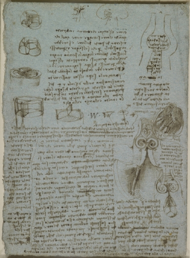
The first accurate drawing of the aortic valve was by Leonardo da Vinci in 1512 (Figure 1). Anatomist Andreas Vesalius (1514-1564), born in Brussels (Belgium), changed Galenic thinking [2]. After Louvain and Paris, he went to Padua (Italy), one of the most prominent universities after Bologna in the Renaissance period [2]. By performing autopsies on humans instead of animals, he discovered Galen’s mistakes. He wrote his key work in 1543, “De Humani Corporis Fabrica” [2-4]. This is regarded as the largest single contribution to medical science [2]. He became so famous after this publication that he immediately became the doctor of Emperor Charles V, as well as of his successor Philip II [2]. However, the concept of circulation had still not been revealed. Building on Vesalius’ work, William Harvey (1578-1657) discovered the property of veins, publishing his findings in his key manuscript “De motu cordis” (1628) [3]. He concluded that blood circulates in a single direction, with the heart as a mechanical pump (5).

Figure 1. Leonardo Da Vinci (Vinci 1452-Amboise 1519)
The Aortic Valve c.1512-13.
Pen and ink on blue paper | 28.3 x 20.4 cm
202_Kuijpers_Figure 1.jpg
Royal Collection Trust / © Her Majesty Queen Elizabeth II 2019

Medical Technology

The Stethoscope

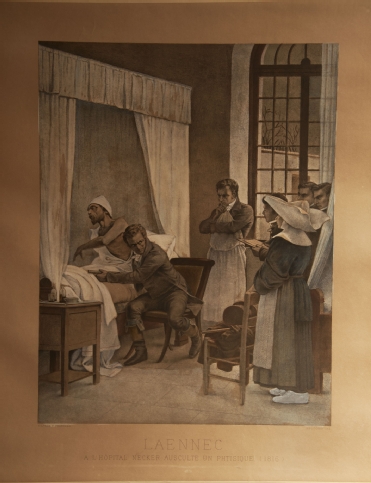
In the early 1800s, a young French physician was called to examine a young woman. At that time, René Laënnec (1781-1826) was chief physician to the Hôpital Necker in Paris and had been studying with Jean-Nicolas Corvisart (1755-1821), personal physician to Napoleon [1]. In those days, social relationships and rules were very strict, and Laënnec was not permitted to lay his ear on the chest of the female patient, so he had to invent something. He developed a simple wooden cylinder about 23 cm (9 inches) long that could be unscrewed for transport. The stethoscope was “born” (Figure 2) [5,6]. Laënnec published his results in his classic treatise “De l’Auscultation Médiate” in 1819 [7]. The first stethoscope was monaural. American born, but with German parents, George Cammann (1804-1863) claimed the invention of adding two earpieces, thus making it binaural in 1852 [5]. But it was the Irish physician and world traveller Arthur Leared (1822-1879) who had already exhibited “a double instrument with gutta percha tubes” at The Great Exhibition in London in 1851 [8].
In 1831, the British physician James Hope was the first to classify murmurs in “A Treatise on the Disease of the Heart and the Great Vessels” [6,9].

Figure 2. Lithograph “Laennec a L’Opital Necker Auscultate un Physique” (1816) from a painting by Chartran, classical French painter (1849-1907). Under the image “D’Apres T Chartran” is engraved on the left, “Heliogravure” on the right.
202_Kuijpers_Figure 2.jpg

Blood Pressure Measurement

French physicist and physiologist Jean Poiseuille (1797-1869, who gave his name to Poiseuille’s law) was very interested in the flow of blood in vessels. In 1828 he presented a mercury manometer device to measure blood pressure [6]. It was the Italian Scipione Riva-Rocci (1863-1937) (hence the denomination RR for blood pressure) who in 1896 introduced the inflatable arm cuff which was coupled to a sphygmograph [6]. Nicolai Korotkoff (1874-1920), a Russian military surgeon, was the first to auscultate the brachial artery sounds in 1905 [6].

X-ray

Another spectacular advance in diagnostic possibilities was the discovery of X-rays in 1895 by the German Wilhelm Conrad Röntgen (1845-1923) [1,5]. In 1908 another German, Morris Simmonds (1855-1925), used X-rays to show valvular calcifications after death for the first time [10]. The American Merrill Sosman (1890-1959), amongst others, diagnosed aortic stenosis (AS) in 1924 in Boston using X-rays in a living person [10].

Catheterisation

The first attempt to enter the heart with a catheter, albeit in a horse, was performed by Frenchman Claude Bernard (1813-1878) in 1844. He managed to reach the left and right ventricles using a catheter via the jugular vein and carotid artery [11]. In 1929, the German Werner Forssmann (1904-1979), at that time working in Eberswalde, had another idea and used himself as a guinea pig; he passed a catheter in his left arm up until the right atrium, using fluoroscopy via a mirror held by a nurse. He then took the stairs to the radiology department to document this on a chest X-ray. Later he decided to become a urologist [6,11].
However, it took until 1950 for the first retrograde left catheterisation to be performed by the American Henry Zimmerman (1915-2007) in Cleveland, as well as Limon Lason in Mexico [11]. Zimmerman was the first to measure left ventricular pressure in 10 patients with aortic regurgitation (AR) [12]. In 1950, the Cuban Rodrigo Bustamente Marcayda was the first to observe AR after injecting contrast in the aorta [12]. In 1953, Swede Sven Ivar Seldinger (1921-1998) developed the percutaneous technique. In 1959, the American F. Mason Sones (1918-1985) was trying to inject contrast dye into the aorta of a young man, but his catheter tipped into the right coronary artery (RCA). Before he could ask to pull back the catheter, a large dose of contrast was already injected, and the RCA was depicted. The patient immediately had an asystole but after coughing his heart rhythm was restored. By serendipity he had just completed the first selective coronary angiography in Cleveland [6,11].
In 1956, Cournand, Forssmann and Richards received the Nobel Prize for their work on heart catheterisation [5].

Echocardiography

The importance of echo reflection was first demonstrated by the Italian Lazzaro Spallanzani (1729-1799). He demonstrated that bats use reflected echoes of inaudible sounds to fly and navigate [13]. Only after the discovery of piezoelectricity in 1880 by Pierre Curie and his brother Jacques did it become possible to create ultrasonic waves [13]. Major advances were bolstered by two wars. Frenchman Paul Langevin (1872-1946) developed Sound Navigation and Ranging (SONAR) in 1915 to detect enemy submarines, followed by the US navy using Radio Detection and Ranging (RADAR) in 1941 to detect planes. Ultrasonic pulse-echo was first used by the Soviet Sergei Sokolov in 1937 to detect flaws in metals [13].
It took several decades before this technology was used in cardiology. The founding father is the Swede Inge Edler (1911-2001). During the late 1940s, Edler, as head of the Cardiovascular Laboratory in Lund, was responsible for preoperative evaluation of (mitral) valve disease for his pioneering heart surgery colleagues Wulff and Sandblom [13]. However, Edler was not happy with his cardiac catheterisation results. He had read a book on RADAR and wondered whether this could be the solution for non-invasive use. Via Jan Cederlund, he came into contact with Carl Hellmut Hertz, son of Nobel Prize Physics winner Gustav Hertz, and nephew of Heinrich Hertz (who gave his name to the unit of frequency). They were lent the first ultrasonic reflectoscope from a shipyard to do some experiments during the weekend. To their fascination, they saw an echo moving back and forth on the oscilloscope screen at a depth of 8-9 cm from the chest wall. Without funding, luckily, they found out that Siemens was building an ultrasonic reflectoscope. Siemens’ boss Gellinek sent the scope to Lund to use for one year; it never went back. After testing and experimenting, they chose 2.5 MHz as the optimal frequency [13]. On 29 October 1953, equipped with the Ultraschall-Impulsgerät, Edler and Hertz recorded the first moving pictures of the heart, thus inaugurating the field of “ultrasound cardiography” [13]. 
Edler and Hertz also attempted transoesophageal echocardiography (TEE) but encountered several difficulties. The credits for the first TEE (performed in Chicago) go to American Leon Frazin in 1976 [13,14]. Two-dimensional echocardiography was introduced in 1974 [6], pulsed Doppler haemodynamics in 1975, and Doppler colour flow in 1982 [6]. Transoesophageal imaging in 1985 made a large contribution to very specific images of cardiac valves [6].
In the USA, Harvey Feigenbaum gave the first course on cardiac ultrasonography in 1968. Feigenbaum is said to have introduced the term “echocardiography” [13].

Aortic Regurgitation (AR)

Galen and Da Vinci understood that the function of the aortic valve was to prevent a reversal of flow [12]. Early descriptions of AR were made in 1706 by London surgeon William Cowper (1666-1709), who also found an enlarged heart on autopsy - “larger than an ox”, hence the name cor bovinum [6,12]. In 1715, French physician Raymond Vieussens (1641-1716) described the collapsing pulse in AR and also confirmed on autopsy an enlarged left ventricle besides the “markedly stretched valves” [6,12]. In Padua in 1761, Italian Giovanni Battista Morgagni (1682-1771) recognised the important haemodynamic consequences of AR [6,12].
In 1832, Englishman James Hope (1801-1841) described a prolonged murmur after the second heart sound [2]. In the same year, Dublin surgeon Sir Dominic Corrigan (1802-1880) provided the classic description of the arterial pulse and also the murmur of AR [6,12]. He noted two other signs - a “bruit de soufflet” over the ascending aorta, carotid and subclavian arteries, as well as a “frémissement” (thrill) over the carotid and subclavian arteries [12]. It was Thomas Watson who, in 1843, linked this pulse, also called pulsus celer, to a water hammer ([2,15]. A water hammer was a Victorian toy in which a tube was half filled with fluid, the remainder being a vacuum. Each time the tube was inverted or shaken, the impact of the fluid at each end would sound like a hammer blow.
In the mid-19th century, Americans who had studied in Paris tried to popularise the use of the stethoscope in the USA. The “American Laënnec” was Austin Flint (1812-1886) [16].
In 1862, he described two patients with AR, each of whom had a diastolic murmur similar to the one heard in mitral stenosis. Flint believed that such a murmur could be produced without any mitral lesions. The regurgitant stream of blood was thrust into the stream leaving the left atrium, thereby setting the mitral leaflets into vibration, which was responsible for the murmur [12,15].
In 1861, Frenchman Paul Louis Duroziez (1826-1897) described an intermittent double murmur over the femoral artery as a sign of AR (“Duroziez’s sign”). The first murmur, resulting from the powerful contraction of the left ventricle, was perceived by pressing the femoral artery 2 cm above the stethoscope; the second murmur (diastolic), believed to be the result of contraction of the arteries in the legs, was heard by pressing the femoral artery 2 cm below the stethoscope [12]. This is not the same as Traube’s sign (Ludwig Traube, 1818-1876, German physician), which is a double tone heard without compressing the femoral artery. The systolic sound, likened to that of a pistol shot, may be due to the sudden distention of the arterial wall [12]. The synchronous nodding of the head with the heart beat was observed in 1900 by Frenchman Delpeuch, who named it “de Musset’s sign” (after a French poet with AR) [12].

Aortic Stenosis (AS)

AS was first described in 1663 in “Opera Medica Universa” by Frenchman Lazare Riviere (1589-1655), based on a patient seen in 1646 [10]. It was also described by the founder of pathological anatomy, Giovanni Battista Morgagni [4,10]. Jean Nicolas Corvisart mentioned an aortic valve “ossified and united so closely that the end of a little finger could scarcely be introduced” [10]. In 1819, Rene Laënnec pointed out that the aortic valve was subject to ossification [6,10].
In the first half of the 20th century, investigators debated on the different causes of AS [10]. A congenital cause of AS was largely ignored; however, the bicuspid aortic valve had already been described in 1844 by British surgeon Sir James Paget (1814-1899) [10].
James Hope provided details of AS auscultation in 1832 [6,10]. Irishman William Stokes (1804-1878) wrote a case on a man who dropped dead from extreme AS [10]. In 1955, Reinhold recorded with phonocardiography a loud early ejection click in a patient with a congenital AS [10].
Besides X-rays, catheterisation was also used, in the beginning via needle punctures. It took years before gradients and left-sided pressures were measured via retrograde arterial catheterisation [10]. In 1950, Limon Lason reported measurements of left ventricular and aortic pressure in AS patients, using a radial artery approach [10].
In 1933, grading of systolic murmurs was introduced in Boston by the Polish born (but living in America from the age of 3) Samuel Levine (1891-1966) [6]. The acoustic principles of cardiovascular sound improved considerably thanks to the work of Rappaport and Sprague in the 1940s [6].

Aetiology

The aetiology of valvular disease in the 19th and first half of the 20th century was mainly about highly prevalent rheumatic fever and gonorrhoea [6]. Infections, such as streptococcal ones, could not be treated as antibiotics had not yet been invented. Doctors only had their stethoscope to examine patients. Improved diagnostic technology yielded other causes of valvular disease. After degenerative calcification, from 1997 the central hypothesis of AS was inflammation of the aortic valve in patients with atherosclerotic risk factors [6,17]. Currently, hallmark features are early atherosclerosis, cell proliferation and osteoblast expression [17].

Valve Surgery and Interventions

In 1913, Frenchman Theodore Tuffier (1857-1929) performed the first operation for AS at the Hôpital de la Pitié in Paris, France. He wanted to incise the stenosed valve but eventually performed a digital dilatation [10]. In the 1920s most interventions were performed on the mitral valve. In 1950, in the Mayo Clinic, American Robert Glover used a dilating instrument on the aortic valve commissurotomy instead of a finger for AS [10].
The next important development in the surgical management of aortic valve disease was the successful implantation of an acrylic ball valve prosthesis in the descending thoracic aorta in a 30-year-old woman with severe AR. This was carried out by Charles Hufnagel in 1952 in Washington DC [10,12,18]. In 1956, Irishman Jeremy Swan (1922-2005, co-inventor of the Swan-Ganz catheter) and Kortz performed the first direct vision commissurotomy for AS, using an ice bath for hypothermia at the Mayo Clinic, Rochester, MN, USA [10].
After the heart-lung machine was designed, Boston surgeon Dwight Harken (1910-1993) was, in 1960, able to implant the first prosthetic valve in the aortic position for AR [12,19].
Walton Lillehei (1918-1999) is regarded as the father of open-heart surgery. He was born in Minneapolis to a Norwegian family [20]. In 1957 he performed the first intracardiac aortic valve replacement with a plastic prosthesis [20]. He was involved in the development of several types of valve, such as the St. Jude valve [20]. During his cardiac surgeries, Lillehei had noted that quite often patients showed conduction disturbances. He was the first to implant an internal cardiac pacemaker, which was at that time connected to an external power source. This gave rise to a lot of practical problems. He called on his electrical engineer Earl Bakken to think of a solution. The rest is history: Medtronic was founded in a garage in Minneapolis in 1958 [20].
In 1960, in Boston, Harken replaced a stenosed aortic valve below the level of the coronary ostia with a caged-ball prosthesis [10,18]. This was at a time when there was great enthusiasm for this type of prosthesis, after an initial report by Albert Starr (physician) and Lowell Edwards (electrical engineer) [18]. This caged-ball type was initially designed for the mitral valve [18].
The first catheter-based approach to balloon aortic valvuloplasty (BAV) was developed by Frenchman Alain Cribier in 1985 [21]. It was the Dane, Henning Rud Andersen who, in 1988,  speculated that, just like a coronary stent, a balloon-expandable valve could be placed, namely transcatheter aortic valve replacement (TAVR) [21]. He could not find a company to develop this, but Cribier with others formed a start-up company. In 2002, he took the credit for the first human percutaneous transcatheter aortic valve implantation (TAVI), performed in Rouen, France [21]. Later, the life of Henning Rud Andersen’s father was saved by the invention of his son, as he regained his life after a TAVI procedure.

Conclusion

Although it is impossible to mention everybody who played a role in the discovery, developments and inventions regarding the aortic valve, this paper provides some insight into the great names involved. Europe played an important role in the development and invention of diagnostic instruments in the early days and since, due to worldwide collaborations, the medical field has progressed. This historical knowledge can inspire us to think beyond imagination, as our predecessors did.

References


  1. Rhodes P, Guthrie DJ, Thomson WAR, Underwood EA, Richardson RG. Encyclopaedia Britannica online. 2017; 25 October. 
  2. Mesquita ET, Souza Junior CV, Ferreira TR. Andreas Vesalius 500 years--A Renaissance that revolutionized cardiovascular knowledge. Rev Bras Cir Cardiovasc. 2015;30:260-5. 
  3. Reichert P. A history of the development of cardiology as a medical specialty. Clin Cardiol. 1978;1:5-15. 
  4. O’Rahilly R. Introduction: General Anatomy. In: Gardner E, Gray DJ, O’Rahilly R, eds. Anatomy: a regional study of human structure. 4th ed. Philadelphia: W.B. Saunders Company; 1975. pp 3-8.
  5. Porter R. The Cambridge illustrated history of medicine. Cambridge: Cambridge University Press; 2009.
  6. Harrington RA, Silverman ME, Wooley CF. A history of the cardiac diseases, and the development of cardiovascular medicine as a specialty. Hurst’s The Heart. 14th ed. New York: McGraw Hill; 2017.
  7. Agnew R. The prelude to stethoscopy: some French, British and Irish contributions in the early nineteenth century. J Med Biogr. 2003;11:135-41. 
  8. Leared A. On the self-adjusting double stethoscope. The Lancet. 1856;68:138. 
  9. Bedford DE. An early account of aortic incompetence by Thomas cuming (1798-1887). Med Hist. 1967;11:398-401. 
  10. Vaslef SN, Roberts WC. Early descriptions of aortic valve stenosis. Am Heart J. 1993;125:1465-74. 
  11. Grossman W. Cardiac catheterization, angiography, and intervention. 5th ed. Philadelphia: Williams and Wilkins; 1996.
  12. Vaslef SN, Roberts WC. Early descriptions of aortic regurgitation. Am Heart J. 1993;125:1475-83. 
  13. Singh S, Goyal A. The origin of echocardiography: a tribute to Inge Edler. Tex Heart Inst J. 2007;34:431-8. 
  14. Frazin LJ. Clearly, with Regard to Transesophageal Echocardiography, "Need Is the Mother of Invention". J Am Soc Echocardiogr. 2018;31:746-7. 
  15. Constant J. Bedside cardiology. Third ed. Boston: Little, Brown and Company; 1985.
  16. Fye WB. Cardiology in 1885. Circulation. 1985;72:21-6. 
  17. Lerman DA, Prasad S, Alotti N. Calcific Aortic Valve Disease: Molecular Mechanisms and Therapeutic Approaches. Eur Cardiol. 2015;10:108-12. 
  18. Effler DB, Favaloro R, Groves LK. Heart Valve Replacement. Clinical Experience. Ann Thorac Surg. 1965;1:4-24. 
  19. Fye B. Caring for the heart: Mayo Clinic and the rise of specialization. Oxford: Oxford University Press; 2015.
  20. Banerjee A. C Walton Lillehei (1918-99): the versatile pioneer of open-heart surgery. J Med Biogr. 2008;16:150-4. 
  21. Harold JG. The evolution of transcatheter aortic valve replacement. Am Coll Cardiol. 2017 July 20.