Σάββατο 8 Φεβρουαρίου 2020

http://mantzalardas.weebly.com/

The Aortic Valve

History in Medicine: The Aortic Valve

e-Journal
This paper on the aortic valve covers five centuries. From Galenus to the first accurate drawing by da Vinci in 1515, it moves on to Harvey who described the cardiac circulation in 1628. This pivotal work was the start of new developments and inventions (stethoscope by Laennec, 1816) that progressively enabled diagnosis and treatment of aortic valve disorders. From first descriptions of aortic stenosis (Riviere, 1663) and regurgitation (Cowper, 1706), via the first clinical diagnostic procedures (Forssmann, catheterisation, 1929; Edler and Hertz, ultrasound cardiography, 1953), the story ends with ground-breaking therapeutic interventions (Hufnagel, prosthesis, 1952, and Cribier, transcatheter aortic valve implantation, 2002).
Valvular Heart Disease

Introduction

Is it possible to write a historical paper on the aortic valve? To modern cardiologists, the aortic valve has always been a fact, with a large range of diagnostic and therapeutic options at our disposition. But how and when was this knowledge built? Who developed the technologies? Standing on the shoulders of our predecessors, we should not take our current knowledge for granted. Insight into how we have come this far will increase our appreciation of all the possibilities we have to treat our patients.

Early History of Anatomy and Physiology

The foundations for anatomy and embryology in the Western world were laid by Aristotle (384 BCE-322 BCE), a pupil of Plato in Athens. By studying the entire world of living things, he is regarded as being the first great biologist [1]. Greek culture shifted to Alexandria, where a famous medical school was established about 300 BCE. Its two best medical teachers were Herophilus, whose manuscripts on anatomy may have been the first of their kind, and Erasistratus, regarded as the founder of physiology. Erasistratus thought that nerves were hollow tubes containing fluid, and that air entered the lungs and heart and was carried through the body in arteries [1]. 
During the early centuries of the Christian era, many Greek doctors moved to Rome. The most famous of them was Galen of Pergamum or Claudius Galenus (129 CE-216 CE) [2]. Galen regarded anatomy as the foundation of medical knowledge and dissected a range of animals, mostly monkeys. He was among the first to describe the valves of the heart. He also saw the structural differences between arteries and veins. One of his most important demonstrations was that arteries carry blood, not air, as had been taught for 400 years [1]. However, circulation was still not understood. Galen believed that nutrients and venous blood went partly through pores in the intraventricular septum, and in the meantime came into contact with pneuma, leading to formation of arterial blood [2].
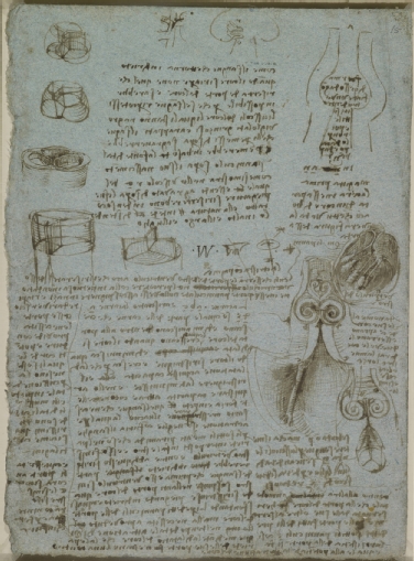
The first accurate drawing of the aortic valve was by Leonardo da Vinci in 1512 (Figure 1). Anatomist Andreas Vesalius (1514-1564), born in Brussels (Belgium), changed Galenic thinking [2]. After Louvain and Paris, he went to Padua (Italy), one of the most prominent universities after Bologna in the Renaissance period [2]. By performing autopsies on humans instead of animals, he discovered Galen’s mistakes. He wrote his key work in 1543, “De Humani Corporis Fabrica” [2-4]. This is regarded as the largest single contribution to medical science [2]. He became so famous after this publication that he immediately became the doctor of Emperor Charles V, as well as of his successor Philip II [2]. However, the concept of circulation had still not been revealed. Building on Vesalius’ work, William Harvey (1578-1657) discovered the property of veins, publishing his findings in his key manuscript “De motu cordis” (1628) [3]. He concluded that blood circulates in a single direction, with the heart as a mechanical pump (5).

Figure 1. Leonardo Da Vinci (Vinci 1452-Amboise 1519)
The Aortic Valve c.1512-13.
Pen and ink on blue paper | 28.3 x 20.4 cm
202_Kuijpers_Figure 1.jpg
Royal Collection Trust / © Her Majesty Queen Elizabeth II 2019

Medical Technology

The Stethoscope

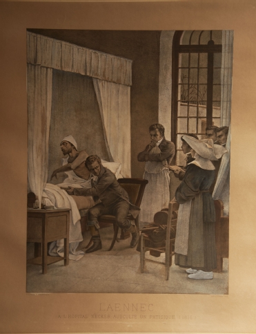
In the early 1800s, a young French physician was called to examine a young woman. At that time, René Laënnec (1781-1826) was chief physician to the Hôpital Necker in Paris and had been studying with Jean-Nicolas Corvisart (1755-1821), personal physician to Napoleon [1]. In those days, social relationships and rules were very strict, and Laënnec was not permitted to lay his ear on the chest of the female patient, so he had to invent something. He developed a simple wooden cylinder about 23 cm (9 inches) long that could be unscrewed for transport. The stethoscope was “born” (Figure 2) [5,6]. Laënnec published his results in his classic treatise “De l’Auscultation Médiate” in 1819 [7]. The first stethoscope was monaural. American born, but with German parents, George Cammann (1804-1863) claimed the invention of adding two earpieces, thus making it binaural in 1852 [5]. But it was the Irish physician and world traveller Arthur Leared (1822-1879) who had already exhibited “a double instrument with gutta percha tubes” at The Great Exhibition in London in 1851 [8].
In 1831, the British physician James Hope was the first to classify murmurs in “A Treatise on the Disease of the Heart and the Great Vessels” [6,9].

Figure 2. Lithograph “Laennec a L’Opital Necker Auscultate un Physique” (1816) from a painting by Chartran, classical French painter (1849-1907). Under the image “D’Apres T Chartran” is engraved on the left, “Heliogravure” on the right.
202_Kuijpers_Figure 2.jpg

Blood Pressure Measurement

French physicist and physiologist Jean Poiseuille (1797-1869, who gave his name to Poiseuille’s law) was very interested in the flow of blood in vessels. In 1828 he presented a mercury manometer device to measure blood pressure [6]. It was the Italian Scipione Riva-Rocci (1863-1937) (hence the denomination RR for blood pressure) who in 1896 introduced the inflatable arm cuff which was coupled to a sphygmograph [6]. Nicolai Korotkoff (1874-1920), a Russian military surgeon, was the first to auscultate the brachial artery sounds in 1905 [6].

X-ray

Another spectacular advance in diagnostic possibilities was the discovery of X-rays in 1895 by the German Wilhelm Conrad Röntgen (1845-1923) [1,5]. In 1908 another German, Morris Simmonds (1855-1925), used X-rays to show valvular calcifications after death for the first time [10]. The American Merrill Sosman (1890-1959), amongst others, diagnosed aortic stenosis (AS) in 1924 in Boston using X-rays in a living person [10].

Catheterisation

The first attempt to enter the heart with a catheter, albeit in a horse, was performed by Frenchman Claude Bernard (1813-1878) in 1844. He managed to reach the left and right ventricles using a catheter via the jugular vein and carotid artery [11]. In 1929, the German Werner Forssmann (1904-1979), at that time working in Eberswalde, had another idea and used himself as a guinea pig; he passed a catheter in his left arm up until the right atrium, using fluoroscopy via a mirror held by a nurse. He then took the stairs to the radiology department to document this on a chest X-ray. Later he decided to become a urologist [6,11].
However, it took until 1950 for the first retrograde left catheterisation to be performed by the American Henry Zimmerman (1915-2007) in Cleveland, as well as Limon Lason in Mexico [11]. Zimmerman was the first to measure left ventricular pressure in 10 patients with aortic regurgitation (AR) [12]. In 1950, the Cuban Rodrigo Bustamente Marcayda was the first to observe AR after injecting contrast in the aorta [12]. In 1953, Swede Sven Ivar Seldinger (1921-1998) developed the percutaneous technique. In 1959, the American F. Mason Sones (1918-1985) was trying to inject contrast dye into the aorta of a young man, but his catheter tipped into the right coronary artery (RCA). Before he could ask to pull back the catheter, a large dose of contrast was already injected, and the RCA was depicted. The patient immediately had an asystole but after coughing his heart rhythm was restored. By serendipity he had just completed the first selective coronary angiography in Cleveland [6,11].
In 1956, Cournand, Forssmann and Richards received the Nobel Prize for their work on heart catheterisation [5].

Echocardiography

The importance of echo reflection was first demonstrated by the Italian Lazzaro Spallanzani (1729-1799). He demonstrated that bats use reflected echoes of inaudible sounds to fly and navigate [13]. Only after the discovery of piezoelectricity in 1880 by Pierre Curie and his brother Jacques did it become possible to create ultrasonic waves [13]. Major advances were bolstered by two wars. Frenchman Paul Langevin (1872-1946) developed Sound Navigation and Ranging (SONAR) in 1915 to detect enemy submarines, followed by the US navy using Radio Detection and Ranging (RADAR) in 1941 to detect planes. Ultrasonic pulse-echo was first used by the Soviet Sergei Sokolov in 1937 to detect flaws in metals [13].
It took several decades before this technology was used in cardiology. The founding father is the Swede Inge Edler (1911-2001). During the late 1940s, Edler, as head of the Cardiovascular Laboratory in Lund, was responsible for preoperative evaluation of (mitral) valve disease for his pioneering heart surgery colleagues Wulff and Sandblom [13]. However, Edler was not happy with his cardiac catheterisation results. He had read a book on RADAR and wondered whether this could be the solution for non-invasive use. Via Jan Cederlund, he came into contact with Carl Hellmut Hertz, son of Nobel Prize Physics winner Gustav Hertz, and nephew of Heinrich Hertz (who gave his name to the unit of frequency). They were lent the first ultrasonic reflectoscope from a shipyard to do some experiments during the weekend. To their fascination, they saw an echo moving back and forth on the oscilloscope screen at a depth of 8-9 cm from the chest wall. Without funding, luckily, they found out that Siemens was building an ultrasonic reflectoscope. Siemens’ boss Gellinek sent the scope to Lund to use for one year; it never went back. After testing and experimenting, they chose 2.5 MHz as the optimal frequency [13]. On 29 October 1953, equipped with the Ultraschall-Impulsgerät, Edler and Hertz recorded the first moving pictures of the heart, thus inaugurating the field of “ultrasound cardiography” [13]. 
Edler and Hertz also attempted transoesophageal echocardiography (TEE) but encountered several difficulties. The credits for the first TEE (performed in Chicago) go to American Leon Frazin in 1976 [13,14]. Two-dimensional echocardiography was introduced in 1974 [6], pulsed Doppler haemodynamics in 1975, and Doppler colour flow in 1982 [6]. Transoesophageal imaging in 1985 made a large contribution to very specific images of cardiac valves [6].
In the USA, Harvey Feigenbaum gave the first course on cardiac ultrasonography in 1968. Feigenbaum is said to have introduced the term “echocardiography” [13].

Aortic Regurgitation (AR)

Galen and Da Vinci understood that the function of the aortic valve was to prevent a reversal of flow [12]. Early descriptions of AR were made in 1706 by London surgeon William Cowper (1666-1709), who also found an enlarged heart on autopsy - “larger than an ox”, hence the name cor bovinum [6,12]. In 1715, French physician Raymond Vieussens (1641-1716) described the collapsing pulse in AR and also confirmed on autopsy an enlarged left ventricle besides the “markedly stretched valves” [6,12]. In Padua in 1761, Italian Giovanni Battista Morgagni (1682-1771) recognised the important haemodynamic consequences of AR [6,12].
In 1832, Englishman James Hope (1801-1841) described a prolonged murmur after the second heart sound [2]. In the same year, Dublin surgeon Sir Dominic Corrigan (1802-1880) provided the classic description of the arterial pulse and also the murmur of AR [6,12]. He noted two other signs - a “bruit de soufflet” over the ascending aorta, carotid and subclavian arteries, as well as a “frémissement” (thrill) over the carotid and subclavian arteries [12]. It was Thomas Watson who, in 1843, linked this pulse, also called pulsus celer, to a water hammer ([2,15]. A water hammer was a Victorian toy in which a tube was half filled with fluid, the remainder being a vacuum. Each time the tube was inverted or shaken, the impact of the fluid at each end would sound like a hammer blow.
In the mid-19th century, Americans who had studied in Paris tried to popularise the use of the stethoscope in the USA. The “American Laënnec” was Austin Flint (1812-1886) [16].
In 1862, he described two patients with AR, each of whom had a diastolic murmur similar to the one heard in mitral stenosis. Flint believed that such a murmur could be produced without any mitral lesions. The regurgitant stream of blood was thrust into the stream leaving the left atrium, thereby setting the mitral leaflets into vibration, which was responsible for the murmur [12,15].
In 1861, Frenchman Paul Louis Duroziez (1826-1897) described an intermittent double murmur over the femoral artery as a sign of AR (“Duroziez’s sign”). The first murmur, resulting from the powerful contraction of the left ventricle, was perceived by pressing the femoral artery 2 cm above the stethoscope; the second murmur (diastolic), believed to be the result of contraction of the arteries in the legs, was heard by pressing the femoral artery 2 cm below the stethoscope [12]. This is not the same as Traube’s sign (Ludwig Traube, 1818-1876, German physician), which is a double tone heard without compressing the femoral artery. The systolic sound, likened to that of a pistol shot, may be due to the sudden distention of the arterial wall [12]. The synchronous nodding of the head with the heart beat was observed in 1900 by Frenchman Delpeuch, who named it “de Musset’s sign” (after a French poet with AR) [12].

Aortic Stenosis (AS)

AS was first described in 1663 in “Opera Medica Universa” by Frenchman Lazare Riviere (1589-1655), based on a patient seen in 1646 [10]. It was also described by the founder of pathological anatomy, Giovanni Battista Morgagni [4,10]. Jean Nicolas Corvisart mentioned an aortic valve “ossified and united so closely that the end of a little finger could scarcely be introduced” [10]. In 1819, Rene Laënnec pointed out that the aortic valve was subject to ossification [6,10].
In the first half of the 20th century, investigators debated on the different causes of AS [10]. A congenital cause of AS was largely ignored; however, the bicuspid aortic valve had already been described in 1844 by British surgeon Sir James Paget (1814-1899) [10].
James Hope provided details of AS auscultation in 1832 [6,10]. Irishman William Stokes (1804-1878) wrote a case on a man who dropped dead from extreme AS [10]. In 1955, Reinhold recorded with phonocardiography a loud early ejection click in a patient with a congenital AS [10].
Besides X-rays, catheterisation was also used, in the beginning via needle punctures. It took years before gradients and left-sided pressures were measured via retrograde arterial catheterisation [10]. In 1950, Limon Lason reported measurements of left ventricular and aortic pressure in AS patients, using a radial artery approach [10].
In 1933, grading of systolic murmurs was introduced in Boston by the Polish born (but living in America from the age of 3) Samuel Levine (1891-1966) [6]. The acoustic principles of cardiovascular sound improved considerably thanks to the work of Rappaport and Sprague in the 1940s [6].

Aetiology

The aetiology of valvular disease in the 19th and first half of the 20th century was mainly about highly prevalent rheumatic fever and gonorrhoea [6]. Infections, such as streptococcal ones, could not be treated as antibiotics had not yet been invented. Doctors only had their stethoscope to examine patients. Improved diagnostic technology yielded other causes of valvular disease. After degenerative calcification, from 1997 the central hypothesis of AS was inflammation of the aortic valve in patients with atherosclerotic risk factors [6,17]. Currently, hallmark features are early atherosclerosis, cell proliferation and osteoblast expression [17].

Valve Surgery and Interventions

In 1913, Frenchman Theodore Tuffier (1857-1929) performed the first operation for AS at the Hôpital de la Pitié in Paris, France. He wanted to incise the stenosed valve but eventually performed a digital dilatation [10]. In the 1920s most interventions were performed on the mitral valve. In 1950, in the Mayo Clinic, American Robert Glover used a dilating instrument on the aortic valve commissurotomy instead of a finger for AS [10].
The next important development in the surgical management of aortic valve disease was the successful implantation of an acrylic ball valve prosthesis in the descending thoracic aorta in a 30-year-old woman with severe AR. This was carried out by Charles Hufnagel in 1952 in Washington DC [10,12,18]. In 1956, Irishman Jeremy Swan (1922-2005, co-inventor of the Swan-Ganz catheter) and Kortz performed the first direct vision commissurotomy for AS, using an ice bath for hypothermia at the Mayo Clinic, Rochester, MN, USA [10].
After the heart-lung machine was designed, Boston surgeon Dwight Harken (1910-1993) was, in 1960, able to implant the first prosthetic valve in the aortic position for AR [12,19].
Walton Lillehei (1918-1999) is regarded as the father of open-heart surgery. He was born in Minneapolis to a Norwegian family [20]. In 1957 he performed the first intracardiac aortic valve replacement with a plastic prosthesis [20]. He was involved in the development of several types of valve, such as the St. Jude valve [20]. During his cardiac surgeries, Lillehei had noted that quite often patients showed conduction disturbances. He was the first to implant an internal cardiac pacemaker, which was at that time connected to an external power source. This gave rise to a lot of practical problems. He called on his electrical engineer Earl Bakken to think of a solution. The rest is history: Medtronic was founded in a garage in Minneapolis in 1958 [20].
In 1960, in Boston, Harken replaced a stenosed aortic valve below the level of the coronary ostia with a caged-ball prosthesis [10,18]. This was at a time when there was great enthusiasm for this type of prosthesis, after an initial report by Albert Starr (physician) and Lowell Edwards (electrical engineer) [18]. This caged-ball type was initially designed for the mitral valve [18].
The first catheter-based approach to balloon aortic valvuloplasty (BAV) was developed by Frenchman Alain Cribier in 1985 [21]. It was the Dane, Henning Rud Andersen who, in 1988,  speculated that, just like a coronary stent, a balloon-expandable valve could be placed, namely transcatheter aortic valve replacement (TAVR) [21]. He could not find a company to develop this, but Cribier with others formed a start-up company. In 2002, he took the credit for the first human percutaneous transcatheter aortic valve implantation (TAVI), performed in Rouen, France [21]. Later, the life of Henning Rud Andersen’s father was saved by the invention of his son, as he regained his life after a TAVI procedure.

Conclusion

Although it is impossible to mention everybody who played a role in the discovery, developments and inventions regarding the aortic valve, this paper provides some insight into the great names involved. Europe played an important role in the development and invention of diagnostic instruments in the early days and since, due to worldwide collaborations, the medical field has progressed. This historical knowledge can inspire us to think beyond imagination, as our predecessors did.

References


  1. Rhodes P, Guthrie DJ, Thomson WAR, Underwood EA, Richardson RG. Encyclopaedia Britannica online. 2017; 25 October. 
  2. Mesquita ET, Souza Junior CV, Ferreira TR. Andreas Vesalius 500 years--A Renaissance that revolutionized cardiovascular knowledge. Rev Bras Cir Cardiovasc. 2015;30:260-5. 
  3. Reichert P. A history of the development of cardiology as a medical specialty. Clin Cardiol. 1978;1:5-15. 
  4. O’Rahilly R. Introduction: General Anatomy. In: Gardner E, Gray DJ, O’Rahilly R, eds. Anatomy: a regional study of human structure. 4th ed. Philadelphia: W.B. Saunders Company; 1975. pp 3-8.
  5. Porter R. The Cambridge illustrated history of medicine. Cambridge: Cambridge University Press; 2009.
  6. Harrington RA, Silverman ME, Wooley CF. A history of the cardiac diseases, and the development of cardiovascular medicine as a specialty. Hurst’s The Heart. 14th ed. New York: McGraw Hill; 2017.
  7. Agnew R. The prelude to stethoscopy: some French, British and Irish contributions in the early nineteenth century. J Med Biogr. 2003;11:135-41. 
  8. Leared A. On the self-adjusting double stethoscope. The Lancet. 1856;68:138. 
  9. Bedford DE. An early account of aortic incompetence by Thomas cuming (1798-1887). Med Hist. 1967;11:398-401. 
  10. Vaslef SN, Roberts WC. Early descriptions of aortic valve stenosis. Am Heart J. 1993;125:1465-74. 
  11. Grossman W. Cardiac catheterization, angiography, and intervention. 5th ed. Philadelphia: Williams and Wilkins; 1996.
  12. Vaslef SN, Roberts WC. Early descriptions of aortic regurgitation. Am Heart J. 1993;125:1475-83. 
  13. Singh S, Goyal A. The origin of echocardiography: a tribute to Inge Edler. Tex Heart Inst J. 2007;34:431-8. 
  14. Frazin LJ. Clearly, with Regard to Transesophageal Echocardiography, "Need Is the Mother of Invention". J Am Soc Echocardiogr. 2018;31:746-7. 
  15. Constant J. Bedside cardiology. Third ed. Boston: Little, Brown and Company; 1985.
  16. Fye WB. Cardiology in 1885. Circulation. 1985;72:21-6. 
  17. Lerman DA, Prasad S, Alotti N. Calcific Aortic Valve Disease: Molecular Mechanisms and Therapeutic Approaches. Eur Cardiol. 2015;10:108-12. 
  18. Effler DB, Favaloro R, Groves LK. Heart Valve Replacement. Clinical Experience. Ann Thorac Surg. 1965;1:4-24. 
  19. Fye B. Caring for the heart: Mayo Clinic and the rise of specialization. Oxford: Oxford University Press; 2015.
  20. Banerjee A. C Walton Lillehei (1918-99): the versatile pioneer of open-heart surgery. J Med Biogr. 2008;16:150-4. 
  21. Harold JG. The evolution of transcatheter aortic valve replacement. Am Coll Cardiol. 2017 July 20.

Επίδραση υπολιπιδαιμικής θεραπείας στη θνητότητα μετά από επέμβαση αορτοστεφανιαίας παράκαμψης επεμβάσεις
Στην παρούσα μετα-ανάλυση αξιολογήθηκε το όφελος στη θνητότητα της περισσότερο έναντι της λιγότερο εντατικής υπολιπιδαιμικής αγωγής στους ασθενείς που υποβλήθηκαν σε αορτοστεφανιαία παράκαμψη. Αξιολογήθηκαν δεδομένα από 7 τυχαιοποιημένες μελέτες με συνολικό αριθμό 10.420 ασθενών που έλαβαν στατίνη ή συνδυασμό στατίνης με εζετιμίμπη ή αναστολέα της PCSK-9 μετά από καρδιοχειρουργική επέμβαση. Βρέθηκε ελάττωση της καρδιαγγειακής θνητότητας κατά 25% και της συνολικής θνητότητας κατά 14% με χορήγηση υψηλής έντασης υπολιπιδαιμικής αγωγής. Τα αποτελέσματα ήταν παρόμοια τόσο σε κλινικά σταθερούς ασθενείς όσο και σε ασθενείς που είχαν υποστεί οξύ στεφανιαίο σύνδρομο. Συμπερασματικά, ασθενείς με ιστορικό αορτοστεφανιαίας παράκαμψης αντλούν σημαντικό κλινικό όφελος στην καρδιαγγειακή και στη συνολική θνητότητα από την εντατική ελάττωση των επιπέδων της LDL-χολ με χορήγηση υψηλής έντασης υπολιπιδαιμικής θεραπείας.


(Alkhalil M.Atherosclerosis. 2020;293:75-78)
Ελληνική Εταιρεία Λιπιδιολογίας,


Σκορ ασβεστίου και θνητότητα σε καπνιστές
Τα καρδιαγγειακά νοσήματα (ΚΑΝ) και ο καρκίνος αποτελούν τα συχνότερα αίτια θανάτου στους καπνιστές. Στην παρούσα μελέτη αξιολογήθηκε η συσχέτιση του σκορ ασβεστίου, όπως αυτό εκτιμάται με αξονική στεφανιογραφία, με τη θνητότητα σε 5.147 καπνιστές. Ο αριθμός των ασβεστωμένων στεφανιαίων αρτηριών χρησιμοποιήθηκε ως ποιοτικός δείκτης διάχυτης αθηρωμάτωσης. Μετά από παρακολούθηση 11,9 ετών καταγράφηκαν 102 θάνατοι από ΚΑΝ, 54 θάνατοι από στεφανιαία νόσο (ΣΝ) και 123 θάνατοι από καρκίνο. Διπλασιασμός του σκορ ασβεστίου φάνηκε να σχετίζεται με αύξηση της συνολικής θνητότητας κατά 10%, της καρδιαγγειακής θνητότητας κατά 15%, της θνητότητας από ΣΝ κατά 26% και της θνητότητας από καρκίνο κατά 6%. Σκορ ασβεστίου ≥ 400 σχετιζόταν με πολλαπλάσιο κίνδυνο θανάτου συγκριτικά με σκορ = 0. Εικόνα διάχυτη αθηρωμάτωσης στην αξονική στεφανιογραφία αύξανε σημαντικά τη συνολική και την καρδιαγγειακή θνητότητα. Οι συγγραφείς καταλήγουν ότι ποιοτικοί και ποσοτικοί δείκτες ασβέστωσης των στεφανιαίων αρτηριών σχετίζονται θετικά με τη θνητότητα από ΚΑΝ και καρκίνο σε καπνιστές .
(Mirbolouk M, Kianoush S, Dardari Z, et al. Atherosclerosis.2019;294:33-40)

Επιμέλεια:

Χ. Μιχαλακέας, Καρδιολόγος,
Διδάκτωρ Πανεπιστημίου Αθηνών,

Επιστημονικός Συνεργάτης Β' Πανεπιστημιακής Καρδιολογικής Κλινικής,

Π.Γ.Ν.«Αττικόν»

Λ. Ραλλίδης, Καθηγητής Καρδιολογίας,
Β΄ Πανεπιστημιακή Καρδιολογική Κλινική,
Π.Γ.Ν. «Αττικόν»

Ι. Λεκάκης, Ομότιμος Καθηγητής Καρδιολογίας ΕΚΠΑ,
Διευθυντής Καρδιολογικού Τομέα,
Κεντρική Κλινική Αθηνών

Τρίτη 28 Ιανουαρίου 2020

Η άσκηση ενδεχομένως μειώνει τον κίνδυνο καρκίνου έως 25%

  • Iatronet
  • 198 προβολές
Η απόφαση-με το Νέο έτος –να αρχίσει να ασκείται κάποιος και να συνεχίσει-μπορεί ενδεχομένως να μειώσει τον κίνδυνο που αντιμετωπίζει για εμφάνιση καρκίνου, σύμφωνα με νέα έρευνα.
Η σωματική δραστηριότητα σύμφωνα με τις συστάσεις, μπορεί να περιορίσει τον κίνδυνο για ορισμένα είδη καρκίνου, έδειξε νέα έρευνα.
Η απόφαση-με το Νέο έτος –να αρχίσει να ασκείται κάποιος και να συνεχίσει-μπορεί ενδεχομένως να μειώσει τον κίνδυνο που αντιμετωπίζει για εμφάνιση καρκίνου, σύμφωνα με νέα έρευνα.
Σε ευρήματα που δημοσιεύτηκαν στο Journal of Clinical Oncology, ερευνητές του National Cancer Institute, της American Cancer Society, και του Harvard T.H. Chan School of Public Health, αναφέρουν ότι άνθρωποι που ασκούνταν σύμφωνα με τις συστάσεις του National Institutes of Health μπόρεσαν να μειώσουν τον κίνδυνο για 7 είδη καρκίνου μέχρι 25%.
Σε αυτά περιλαμβάνονταν κοινά είδη, όπως του παχέος εντέρου και του μαστού, του ενδομητρίου, των νεφρών, του ήπατος, μυέλωμα και μη Hodgkin λέμφωμα.
Η Alpa Patel, δήλωσε ότι οδηγίες για τη σωματική άσκηση έχουν βασιστεί σε μεγάλο βαθμό στην επίδραση που έχουν στις χρόνιες νόσους, όπως η καρδιαγγειακή νόσος και ο διαβήτης.
Αυτά τα στοιχεία δίνουν ισχυρές ενδείξεις ότι αυτά τα συνιστώμενα επίπεδα είναι σημαντικά και για την πρόληψη του καρκίνου.
Οι ερευνητές ανέλυσαν 9 έρευνες σε 750.000 ενηλίκους συμμετέχοντες και εξέτασαν τη σχέση άσκησης και εμφάνισης 15 ειδών καρκίνου.
Ανακάλυψαν ότι η άσκηση σύμφωνα με τις συστάσεις του NIH- 7,5 έως 15 MET (Metabolic Equivalent Units)  ώρες την εβδομάδα συνδεόταν με σημαντικά χαμηλότερο κίνδυνο για 7 από τα 15 είδη καρκίνου που μελετήθηκαν-με τη μείωση να αυξάνει σε συνάρτηση με περισσότερες MET ώρες.
Για παράδειγμα, η άσκηση συνδεόταν με χαμηλότερο κίνδυνο για καρκίνο στο παχύ έντερο στους άντρες- 8% για 7,5 MET ώρες/εβδομάδα και 14% για  15 MET ώρες/εβδομάδα—και στον καρκίνο μαστού στις γυναίκες -- 6 % για 7,5 MET ώρες/εβδομάδα και 10% για 15 MET ώρες/εβδομάδα. Ο ατομικός κίνδυνος για καρκίνο του ενδομητρίου μειώθηκε κατά 10% έως 18% ενώ για τους νεφρούς, κατά 11 έως 17%.
Ο κίνδυνος για μυέλωμα ήταν μειωμένος κατά 14% έως 19 %, ενώ για το λέμφωμα μη -Hodgkin στις γυναίκες ήταν μειωμένος κατά 11% έως 18%. Η άσκηση είχε την πιο βαθιά επίδραση στον κίνδυνο καρκίνου του ήπατος μειώνοντάς τον κατά  18% έως 27 %.

Δευτέρα 27 Ιανουαρίου 2020

διαβήτης με καρδιακή ανεπάρκεια

'Ερευνα συνδέει τον διαβήτη με την καρδιακή ανεπάρκεια

  • Iatronet
Στην έρευνα που έγινε σε πληθυσμό, ο κίνδυνος καρδιακής ανεπάρκειας ήταν σχεδόν διπλάσιος σε ανθρώπους με διαβήτη έναντι υγιών ανθρώπων.
Ερευνητές του Mayo Clinic πιστεύουν τώρα ότι άνθρωποι με διαβήτη έχουν αυξημένο κίνδυνο για καρδιακή ανεπάρκεια.
Σε ευρήματα που δημοσιευτήκαν στο  Mayo Clinic Proceedings, ερευνητές εντόπισαν ότι ο διαβήτης έχει ανεξάρτητο παράγοντα κινδύνου για εμφάνιση καρδιακής ανεπάρκειας.
Στην έρευνα που έγινε σε πληθυσμό, ο κίνδυνος καρδιακής ανεπάρκειας ήταν σχεδόν διπλάσιος σε ανθρώπους με διαβήτη έναντι υγιών ανθρώπων.
Έχει υποδειχτεί ότι η καρδιακή ανεπάρκεια σε ανθρώπους με διαβήτη μπορεί ενδεχομένως να είναι αποτέλεσμα άλλης πάθησης όπως υπέρτασης ή στεφανιαίας νόσου. Ωστόσο δεν είναι πάντα έτσι.
Οι ερευνητές αξιολόγησαν τη μακροπρόθεσμη επίδραση του διαβήτη στην καρδιακή ανεπάρκεια και εξέτασαν τον κίνδυνο θανάτου.
Από αρχικό δείγμα 2.042 rανθρώπων επέλεξαν 116 ανθρώπους με διαβήτη για την έρευνα και 232 υγιείς. Σε διάστημα 10 ετών το 21% όσων είχαν διαβήτη που εμφάνισαν καρδιακή ανεπάρκεια την εμφάνισαν ανεξάρτητα από άλλα αίτια.
Συγκριτικά, το 12% του δείγματος που δεν είχε διαβήτη εμφάνισε καρδιακή ανεπάρκεια. Οι ερευνητές δεν ανακάλυψαν διαφορά στους θανάτους από καρδιά, στην καρδιακή προσβολή και στο εγκεφαλικό στις 2 ομάδες.
Οι ερευνητές πιστεύουν ότι το εύρημα στηρίζει την έννοια της διαβητικής μυοκαρδιοπάθειας.

Σάββατο 25 Ιανουαρίου 2020

Θρόμβωση και καρκίνος

Θρόμβωση και καρκίνος
 Μια “αθόρυβη” πρόκληση για ιατρούς και ασθενείς της Μαρίας Λυσάνδρου
 Ο Δρ. Ιωάννης Μπουκοβίνας, παθολόγος-ογκολόγος, πρόεδρος της ΕΟΠΕ (Ένωσης Παθολόγων Ογκολόγων Ελλάδας) 23 # 42 / Δεκέμβριος 2019

 Η θρόμβωση φέρεται να επηρεάζει έναν στους πέντε ογκολογικούς ασθενείς. Ποιοι είναι οι βασικότεροι παράγοντες που αυξάνουν τις πιθανότητες ανάπτυξης θρόμβων στους πάσχοντες από καρκίνο; Πράγματι, οι ασθενείς με καρκίνο βρίσκονται σε μεγαλύτερο κίνδυνο για εμφάνιση της θρόμβωσης από τον γενικό πληθυσμό, και είναι σημαντικό να γνωρίζουν ότι η πιθανότητα εμφάνισης θρόμβωσης σχετιζόμενης με τον καρκίνο είναι μεγαλύτερη, ειδικά τους πρώτους μήνες μετά τη διάγνωση της κακοήθειας. Σε αυτό συνεισφέρουν παράγοντες που έχουν σχέση με τον ίδιο τον ασθενή, όπως για παράδειγμα το να έχει ιστορικό θρόμβωσης, προχωρημένη ηλικία, παχυσαρκία, να βρίσκεται σε παρατεταμένη ακινησία ή να έχει πιθανές συνυπάρχουσες παθήσεις. Επιβαρυντικοί, επίσης, μπορεί να είναι παράγοντες που συνδέονται με τον τύπο και το στάδιο του καρκίνου. Παραδείγματος χάριν, οι ασθενείς με εντόπιση της νόσου στο πάγκρεας, στον στόμαχο, στους πνεύμονες, στις ωοθήκες ή στην ουροδόχο κύστη, διατρέχουν μεγαλύτερο κίνδυνο για εμφάνιση θρόμβωσης. Ενώ, αν ο καρκίνος είναι σε προχωρημένο ή μεταστατικό στάδιο, ο κίνδυνος είναι ακόμη μεγαλύτερος. Τέλος, το είδος της ογκολογικής θεραπείας που θα επιλεγεί, όπως κάποιες χημειοθεραπείες ή η ακτινοθεραπεία, αλλά και οι χειρουργικές επεμβάσεις στις οποίες πιθανώς να υποβληθούν οι ασθενείς, παρόλο που είναι απαραίτητες για τη μάχη με τον καρκίνο, είναι παράγοντες που αυξάνουν τον κίνδυνο για θρόμβωση. Ποια μέτρα πρόληψης μπορεί να εφαρμόσει κανείς (και ιδιαίτερα ένας ογκολογικός ασθενής), προκειμένου να ελαττώσει τις πιθανότητες εμφάνισης θρόμβωσης; Η γνώση είναι δύναμη και σε αυτόν τον τομέα. Είναι σημαντικό για έναν ασθενή να γνωρίζει για το ενδεχόμενο εμφάνισης της φλεβικής θρόμβωσης κατά τη διάρκεια της πορείας της νόσου του, να μπορεί να αναγνωρίσει τυχόν προειδοποιητικά σημάδια και να τα αναφέρει εγκαίρως στον γιατρό του. Η αξιολόγηση των επιβαρυντικών παραγόντων και η εκτίμηση του κινδύνου για φλεβική θρόμβωση από τον Ογκολόγο είναι δυναμική, ιδιαίτερα κατά τη διάρκεια της θεραπείας του για τη νόσο, και δύναται να οδηγήσει ακόμη και σε φαρμακολογική προφύλαξη των ασθενών με –τα κατάλληλα για τον κάθε ασθενή ξεχωριστά– αντιπηκτικά φάρμακα. Εκ των ων ουκ άνευ θεωρείται η εκπαίδευση και των ίδιων των ιατρών, σχετικά με την έγκαιρη διάγνωση και αντιμετώπιση περιστατικών που αφορούν στη σχέση θρόμβωσης και καρκίνου.

Σε τι επίπεδο θεωρείτε ότι βρισκόμαστε στην Ελλάδα από πλευράς κατάρτισης των ιατρών, ώστε να αντιμετωπίζουν αποτελεσματικά τέτοια περιστατικά, αλλά και να διαγιγνώσκουν την ανάγκη συνεργασίας, σε κάποιες περιπτώσεις, με ένα συνάδελφο άλλης ειδικότητας;

 Η Ευρωπαϊκή Εταιρεία Ογκολόγων Παθολόγων προσφέρει "μόρια εκπαίδευσης" στους ειδικούς που εντρυφούν στο θέμα της Θρόμβωσης για τους ασθενείς με καρκίνο, και γνωρίζω πως αρκετοί από εμάς τα έχουν κομίσει. Εμείς, ως Ελληνική Ογκολογική Εταιρεία, έχουμε συμπεριλάβει το θέμα της Θρόμβωσης στην εκπαίδευση των Νέων Συναδέλφων, και στα συνέδριά μας αποτελεί συχνά θέμα ενημέρωσης και συζήτησης. Οπότε, εκτιμώ πως η ενημέρωση των ειδικών είναι σε καλό επίπεδο, αλλά δεν προτείνεται εφησυχασμός. Ωστόσο, η πρόκληση της επιστήμης μας είναι η συνεχιζόμενη ενημέρωση και εκπαίδευση όχι μόνο για την θρόμβωση, αλλά για όλα τα νεότερα δεδομένα που προκύπτουν και μπορούν να ενδυναμώσουν την φροντίδα μας για τον ασθενή. Πιστεύουμε πως ο ασθενής χρειάζεται μια ολιστική προσέγγιση. Και ο κίνδυνος για τη θρόμβωση είναι ένας από τους παράγοντες που αξιολογούνται και, αν χρειαστεί, και σε συνεργασία με άλλες ειδικότητες. Ένα πολύ σημαντικό βήμα ως προς τη διαχείριση της σχετικής με τον καρκίνο θρόμβωσης σημειώθηκε στο πλαίσιο του φετινού Πανευρωπαϊκού Ογκολογικού Συνεδρίου - ESMO congress, το οποίο πραγματοποιήθηκε στη Βαρκελώνη, από τις 27 Σεπτεμβρίου έως την 1η Οκτωβρίου. Για πρώτη φορά στο συγκεκριμένο συνέδριο, οργανώθηκε σχετικό δορυφορικό συμπόσιο, στο οποίο προήδρευσε ο Δρ. Πάρις Κοσμίδης, παθολόγος-ογκολόγος, διευθυντής Β’ Παθολογικής-Ογκολογικής Κλινικής ΔΘΚΑ "Υγεία". Ο ίδιος αναλαμβάνει να μας διαφωτίσει περαιτέρω. "Η θρόμβωση και η εμβολή είναι νοσηρές οντότητες, συνυφασμένες με τον καρκίνο. Συνυπάρχουν, προηγούνται ή έπονται του καρκίνου. Το 20% των ασθενών με καρκίνο θα αναπτύξουν, σε κάποια φάση της αρρώ- 24 top stories O Δρ. Πάρις Κοσμίδης, παθολόγος-ογκολόγος, διευθυντής Β’ ΠαθολογικήςΟγκολογικής Κλινικής ΔΘΚΑ "Υγεία". στιας τους, θρομβοεμβολική νόσο, αποτελώντας μάλιστα συχνό αίτιο θανάτου", αναφέρει ο κ. Κοσμίδης και σημειώνει ότι υπάρχουν διάφοροι μηχανισμοί στον οργανισμό που εξηγούν αυτήν τη σχέση μεταξύ θρόμβωσης και καρκίνου, που διαφέρουν ανάλογα με τον τύπο του καρκίνου: Σε γενικές γραμμές, απελευθερώνονται από τον καρκίνο ουσίες που διευκολύνουν και προάγουν την υπερπηκτικότητα του αίματος. Το φαινόμενο αυτό, σε συνδυασμό με επηρεασμένα αγγεία, χειρουργεία, αντικαρκινικά φάρμακα, ακτινοθεραπείες, δυσκινησία των ασθενών κ.λπ., συμβάλλει στη δημιουργία της θρομβοεμβολικής νόσου. Είναι αυτονόητο ότι παχύσαρκα, ευτραφή άτομα έχουν μεγαλύτερο κίνδυνο, όπως και για καρκίνο του γαστρεντερικού συστήματος, πνεύμονος, γυναικολογικών οργάνων κ.λπ. "Από τα αντικαρκινικά φάρμακα, η πλατίνα, οι ορμόνες, αλλά και πρόσφατα οι ανοσοθεραπείες, ενέχονται στη δημιουργία θρόμβωσης", προσθέτει ο ειδικός. Τι ενδείκνυται, λοιπόν, σε αυτές τις περιπτώσεις; "Επειδή οι ασθενείς που εμφανίζουν θρομβοεμβολική νόσο έχουν μεγαλύτερη πιθανότητα να πεθάνουν, σε σύγκριση με τους υπόλοιπους, η θεραπεία πρέπει να είναι άμεση και αποτελεσματική. Η χορήγηση χαμηλού μοριακού βάρους Ηπαρίνης είναι η επιβεβλημένη θεραπεία. Η διάρκεια της θεραπείας μπορεί να κρατήσει πέραν των 6 μηνών, κατά την κρίση του θεράποντος ιατρού. Προληπτικά, ο γιατρός έχει δυνατότητα να χορηγήσει την Ηπαρίνη χαμηλού μοριακού βάρους σε επιλεγμένους ασθενείς με καρκίνο, π.χ. αμέσως μετά το χειρουργείο, σε κλινήρεις ασθενείς, σε παχύσαρκους. Επίσης, σε ορισμένες μορφές καρκίνου, όπως πνεύμονος, γαστρεντερικού, γυναικολογικών οργάνων, ακολουθώντας συγκεκριμένους αλγόριθμους. Ακόμη, σε ασθενείς με καρκίνο που αρχίζουν χημειοθεραπεία με πλατίνα, η προφυλακτική αντιπηκτική θεραπεία έχει υπό προϋποθέσεις ένδειξη. Επειδή η Ηπαρίνη χαμηλού μοριακού βάρους γίνεται υποδορίως και αυτό προκαλεί κάποια δυσφορία στους ασθενείς, έχει γίνει προσπάθεια για χρησιμοποίηση από του στόματος δισκία αντιπηκτικά. Ωστόσο, στους ασθενείς με καρκίνο δεν έχουν ακόμη βρει εφαρμογή, διότι προκαλούν αιμορραγίες, ιδίως σε όγκους του γαστρεντερικού, αλλά και αλληλοεπιδράσεις με άλλα αντικαρκινικά φάρμακα". Το συμπέρασμα, σύμφωνα με τον κ. Κοσμίδη, είναι ξεκάθαρο: "Η θρόμβωση αποτελεί μια συχνή και επικίνδυνη νοσηρότητα σε ασθενείς με καρκίνο, και η θεραπευτική ή προληπτική της αντιμετώπιση είναι επιβεβλημένη ή επιθυμητή. Πιστεύω ότι οι ογκολογικοί ασθενείς δεν είναι επαρκώς ενημερωμένοι, γι’ αυτό και πρέπει οι γιατροί τους να τους προσφέρουν αυτή τη γνώση. Οι Ογκολόγοι είναι επαρκώς ενημερωμένοι, αλλά υποεκτιμούν τον κίνδυνο". *
www.gateofthrombs.gr

Stable coronary disease with atrial fibrillation

Stable coronary disease with atrial fibrillation: anticoagulants alone or in combination with an antiplatelet agent?

In patients with atrial fibrillation (AF) and stable coronary disease (not requiring revascularisation or revascularised for > 1 year), treatment combining a direct oral anticoagulant (DOAC; rivaroxaban) with an antiplatelet agent (APA) has, compared to a DOAC alone, a risk/benefit ratio which is:
  • A
    Unfavourable
  • B
    Comparable
  • C
    Favourableς, η σωστή απάντηση είναι η εξής Unfavourable
Περισσότερα
An open-label randomised controlled trial included 2,200 Japanese subjects of this type and was stopped early due to increased overall mortality in the DOAC + APA (aspirin or P2Y12 inhibitor) arm, compared to the DOAC alone (3.37% vs. 1.85% per patient-year, respectively), essentially due to an increased rate of major bleeding (2.76% vs. 1.62% per patient-year). The combined treatment was more effective than the monotherapy (composite endpoint of cardiovascular events and mortality), but the difference was non-inferior based on a pre-defined threshold.
Source: Antithrombotic Therapy for Atrial Fibrillation with Stable Coronary Disease

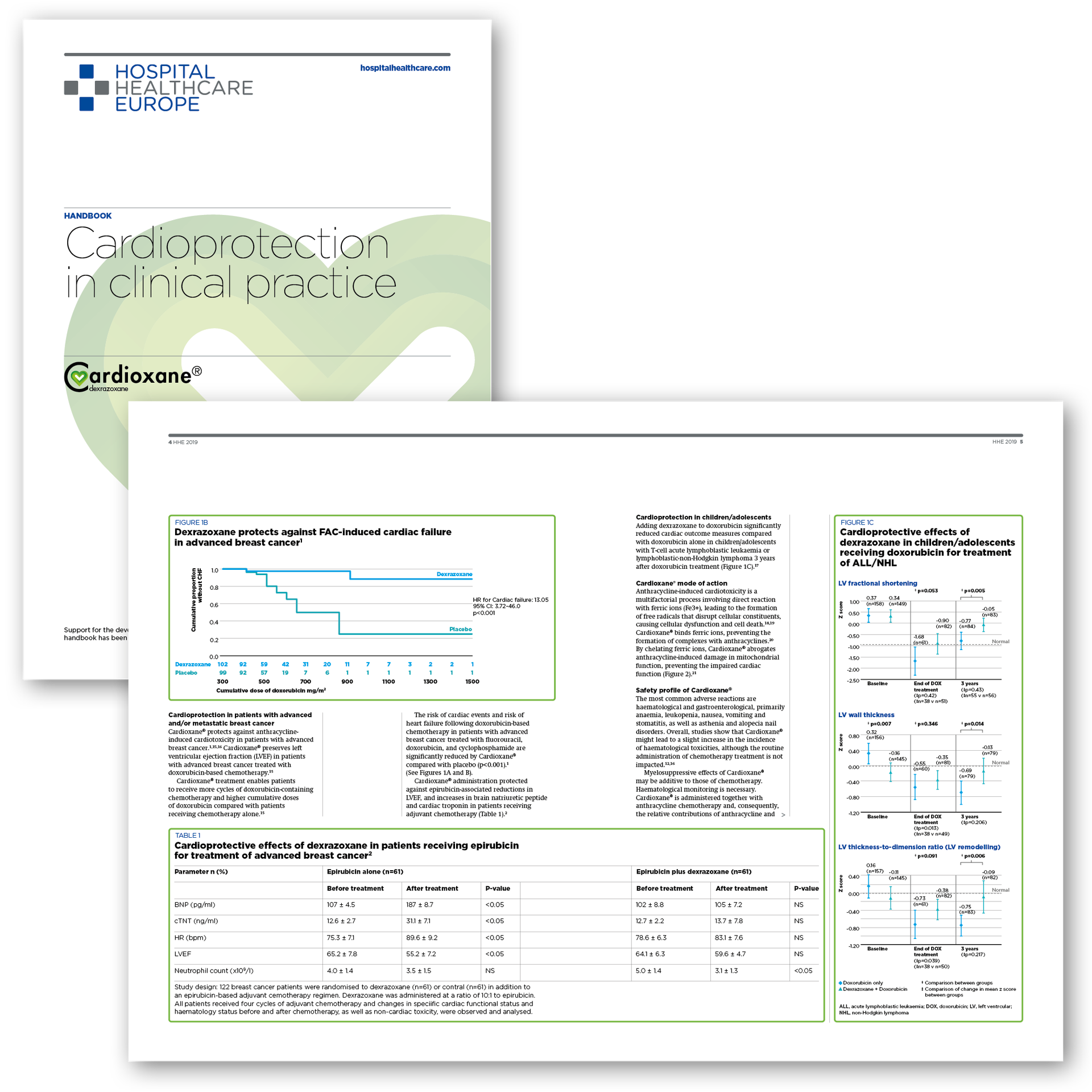
cardioprotection against the toxic effects of anthracyclines

Cardioxane® is a cardioprotective treatment option which is indicated in adults for the prevention of chronic cumulative cardiotoxicity caused by anthracycline use in advanced and/or metastatic breast cancer patients who have received a prior cumulative dose of 300mg/m2 of doxorubicin or a prior cumulative dose of 540mg/m2 of epirubicin when further anthracycline treatment is required.

Hospital Healthcare Europe
I'm an image
I'm an image

Τρίτη 14 Ιανουαρίου 2020

Ο ΡΟΛΟΣ ΤΗΣ ΥΠΕΡΗΧΟΚΑΡΔΙΟΓΡΑΦΙΑΣ ΣΤΗΝ ANTIMETΩΠΙΣΗ ΤΗΣ ΟΞΕΙΑΣ ΠΝΕΥΜΟΝΙΚΗΣ ΕΜΒΟΛΗΣ
 Eπιμέλεια Μ. Μπότης
ΠΕΡΙΛΗΨΗ Ο ρόλος της υπερηχοκαρδιογραφίας στην οξεία πνευμονική εμβολή (ΠΕ) παραμένει ασαφώς καθορισμένος. Είναι μια εξέταση που δεν μπορεί να την διαγνώσει πλήρως και δεν έχει προγνωστική αξία σε ασθενείς με χαμηλού κινδύνου πνευμονική εμβολή, που δεν διαθέτουν άλλα στοιχεία δυσλειτουργίας δεξιάς κοιλίας (ΔΚ). Ωστόσο, πιθανώς να παρέχει προγνωστικές πληροφορίες σε ασθενείς υψηλότερου κινδύνου, όπως επίσης μπορεί να βοηθήσει στην διάκριση οξείας και χρόνιας πνευμονικής εμβολής. Φαίνεται πως συγκεκριμένοι δείκτες μυοκαρδιακής δυσλειτουργίας έχουν την δυνατότητα να παρέχουν καλύτερη προγνωστική αξία από τα υπάρχοντα μοντέλα κινδύνου. Μέχρι όμως να μελετηθούν εντατικά σε προοπτικές μελέτες, η θέση της υπερηχοκαρδιογραφίας σε θεραπευτικό αλλά και οικονομικό επίπεδο παραμένει ασαφώς καθορισμένη. ΕΙΣΑΓΩΓΗ Η έκβαση της πνευμονικής εμβολής βελτιώνεται σταθερά τις τελευταίες δύο δεκαετίες. Η θνητότητα οποιασδήποτε αιτιολογίας αυτή την στιγμή βρίσκεται στο 5-7%, πιθανώς λόγω της βελτιωμένης διαστρωμάτωσης κινδύνου και της χρήσης αποτελεσματικότερων τεχνικών, όπως η θρομβόλυση. Ωστόσο, ανάμεσα στους ασθενείς που παρουσιάζονται με αιμοδυναμική αστάθεια, καταπληξία ή καρδιακή ανακοπή, η θνητότητα 30 ημερών ανεξάρτητα αιτιολογίας παραμένει στο 14-22%. Αντίστοιχα, οι ασθενείς χαμηλού κινδύνου, που αποτελούν ένα ποσοστό 20 με 50% επί του συνόλου, έχουν στο ίδιο διάστημα θνητότητα μικρότερη από 1%. Με τόσο μεγάλες διαφορές λοιπόν στην έκβαση και δεδομένων των εξελισσόμενων θεραπευτικών επιλογών, η διαστρωμάτωση κινδύνου στην ΠΕ είναι πιο επίκαιρη από ποτέ. Η δυσλειτουργία της ΔΚ λόγω της οξείας ΠΕ παραμένει πηγή νοσηρότητας και θνητότητας. Οξεία δυσλειτουργία και βλάβη της ΔΚ (άνοδος των καρδιακών ενζύμων ή/και απεικονιστικά ευρήματα), σε διάφορους βαθμούς, ανιχνεύεται τουλάχιστον στο ήμισυ των ασθενών που νοσηλεύονται λόγω οξείας ΠΕ. Υπάρχουν πολλές μελέτες που έχουν εξετάσει τον ρόλο της υπερηχοκαρδιογραφίας στην διαστρωμάτωση κινδύνου στην ΠΕ. Ωστόσο, ελάχιστες μελέτες έχουν επιχειρήσει να προσαρμόσουν στα υπερηχοκαρδιογραφικά ευρήματα τους δείκτες δυσλειτουργίας ή βλάβης της ΔΚ και μόνο πρόσφατα τα μοντέλα κινδύνου ενσωμάτωσαν καρδιακούς βιοδείκτες και ή/και ευρήματα δυσλειτουργίας ΔΚ, μέσω αξονικής τομογραφίας. Επίσης, οι κατευθυντήριες οδηγίες της European Society of Cardiology (ESC) και του American College of Chest Physicians συμβούλεψαν κατά της καθιέρωσης της υπερηχοκαρδιογραφίας στην οξεία ΠΕ. Οι κατευθυντήριες οδηγίες δεν είναι δυνατό να ενσωματώσουν όλα τα πιθανά σενάρια στα οποία η υπερηχοκαρδιογραφία μπορεί να βοηθήσει στην λήψη κλινικών αποφάσεων. Μια κριτική ανασκόπηση της βιβλιογραφίας μπορεί να βοηθήσει στην λεπτή απόφαση διεξαγωγής υπερηχοκαρδιογραφίας στην οξεία ΠΕ. Σε αυτή την ανασκόπηση, θα εξετάσουμε την υπάρχουσα βιβλιογραφία για τον ρόλο της υπερηχοκαρδιογραφίας στην διάγνωση και στην διαστρωμάτωση κινδύνου στην οξεία ΠΕ. Επίσης, θα συζητηθούν πιθανοί υπερηχοκαρδιογραφικοί δείκτες κινδύνου αλλά και ο ρόλος της υπερηχοκαρδιογραφίας στις θεραπευτικές αποφάσεις και στην βελτιστοποίηση της αντιμετώπισης αυτών των ασθενών. Η ΥΠΕΡΗΧΟΚΑΡΔΙΟΓΡΑΦΙΑ ΚΑΙ Η ΔΙΑΓΝΩΣΗ ΟΞΕΙΑΣ ΠΕ Οι διαγνωστικές επιδόσεις της αξονικής αγγειογραφίας πολλαπλών τομών (MDCT) την έχουν καταστήσει ως εξέταση εκλογής στην διάγνωση της οξείας ΠΕ. Η υπερηχοκαρδιογραφία, χάρις στην μη επεμβατική φύση της, την διαθεσιμότητα και την δυνατότητα να πραγματοποιηθεί στην κλίνη του ασθενούς, φαίνεται ως μια αξιόλογη εναλλακτική. Πράγματι, οι ερευνητές επιχείρησαν επανειλημμένα να προσδιορίσουν τον ρόλο τη στην διάγνωση της οξείας ΠΕ. Σε μία πρόσφατη συστηματική ανασκόπηση και μετα-ανάλυση, προσδιορίστηκαν 22 μελέτες που διερεύνησαν την απόδοση της υπερηχοκαρδιογραφίας στην πιθανή οξεία ΠΕ. Πολλές απεικονιστικές τεχνικές χρησιμοποιήθηκαν ως «gold standards”, όπως η MDCT, το σπινθηρογράφημα αερισμού-αιμάτωσης, το χειρουργείο ή η αυτοψία. Στο σύνολο του πληθυσμού που μελετήθηκε, το ποσοστό οξείας πνευμονικής εμβολής ήταν 40.8%. Μόνο η αυξημένη τελοδιαστολική διάμετρος της ΔΚ είχε ευαισθησία 80% για την οξεία ΠΕ (95% CI 61% με 92%), οδηγώντας τους συγγραφείς στο συμπέρασμα πως η υπερηχοκαρδιογραφία υπολείπεται ευαισθησίας για τον αποκλεισμό της ΠΕ. Δυνιτικά χρήσιμα ευρήματα στην διάγνωση επι υποψίας οξείας ΠΕ Εύρημα Pooled Specifity (citation) Estimated positive likehood ratio Estimated negative likehood ratio Θρόμβος δεξιών κοιλοτήτων 0.99 (95% CI 0.96 to 1.0) 5.0 0.96 Σημείο McConnell 0.97 (95% CI 0.95 to 0.99) 7.3 0.80 Παράδοξη κινητικότητα μεσοκοιλιακού διαφράγματος 0.95 (95% CI 0.93 to 0.97) 5.2 0.78 Υποκινησία ελεύθερου τοιχώματος ΔΚ 0.91 (95% CI 0.88 to 0.94) 4.2 0.68 Αυξημένη τελοδιαστολική διάμετρος ΔΚ 0.80 (95% CI 0.61 to 0.92) 4.0 0.25 Συνολική εντύπωση strain ΔΚ 0.83 (95% CI 0.74 to 0.90) 3.1 0.57 Δύο ευρήματα αυτής της μετα-ανάλυσης χρήζουν περαιτέρω αξιολόγησης. Πρώτον, σε πιθανή ΠΕ, υπάρχουν ορισμένα σημεία με υψηλή ειδικότητα: ύπαρξη θρόμβου στις δεξιές κοιλότητες, σημείο McConnell, παράδοξη κινητικότητα μεσοκοιλιακού διαφράγματος και υποκινησίες του ελεύθερου τοιχώματος της ΔΚ. Δεύτερον, η συνολική υπερηχοκαρδιογραφική εντύπωση της δυσλειτουργίας της ΔΚ, ένα μη επαρκώς τεκμηριωμένο αλλά συχνό εύρημα, έχει positive likehood ratio 3.12 εάν τίθεται η υποψία ΠΕ. Ασθενείς με αιμοδυναμική αστάθεια ή καρδιακή ανακοπή μπορεί επίσης να επωφεληθούν από την υπερηχοκαρδιογραφία όταν πιθανολογείται ΠΕ και η «gold standard” απεικόνιση δεν είναι διαθέσιμη. (Στην εικόνα 1, ασθενής υποβλήθηκε σε υπέρηχο για εκτίμηση κατόπιν καρδιαγγειακού συμβάντος. Παρατηρήθηκε σημείο McConnell. Η εικόνα Α λήφθηκε κατά την διαστολή και η εικόνα Β κατά την συστολή. Παρατηρείστε πως μόνο η κορυφή εμφανίζει αξιοσημείωτη κινητικότητα. Στην συνέχεια, διαγνώστηκε οξεία ΠΕ μέσω MDCT. Στην εικόνα C φαίνονται μεσοσυστολικά frames πριν (Εικόνα C) και αφού (Εικόνα D) υπέστη αιμοδυναμικά σημαντική πνευμονική εμβολή που οδήγησε σε καρδιακή ανακοπή. Παρατηρείστε την διάταση της ΔΚ και την παράδοξη κινητικότητα του μεσοκοιλιακού διαφράγματος. Η γεωμετρία της ΑΚ άλλαξε από στρογγυλή σε σχήμα D κατόπιν της οξείας ΠΕ, κάτι που υποδεικνύεται μέσω αυτών των ευρημάτων και επιβεβαιώθηκε μέσω MDCT). Η υπερηχοκαρδιογραφία έχει επίσης θέση στην εγκυμοσύνη και άλλες καταστάσεις που πρέπει να αποφευχθεί η έκθεση σε ακτινοβολία. Ωστόσο, συνίσταται η πλήρης διαγνωστική εκτίμηση θρομβοεμβολικής νόσου, πχ μέσω υπερηχοκαρδιογραφίας φλεβών. Η υπερηχοκαρδιογραφία συνίσταται επίσης για να επιβεβαιωθεί η δυσλειτουργία ΔΚ ως το αίτιο υπότασης σε ασθενείς με γνωστή ή πιθανή ΠΕ, αν και η σύσταση βασίζεται κυρίως σε μη τυχαιοποιημένες μελέτες και άποψη ειδικών. Είναι σημαντικό ότι η ερμηνεία των ευρημάτων σε τέτοια σενάρια από Εικόνα 1 μη-καρδιολόγους έχει υψηλή ακρίβεια και επαναληψιμότητα, επεκτείνοντας τον ρόλο της υπερηχοκαρδιογραφίας στην ορθή διάγνωση.

Γουδέβενος Ιωάννης